Loading
PDBj
MenuPDBj@FacebookPDBj@TwitterPDBj@YouTubewwPDB FoundationwwPDB
RCSB PDBPDBeBMRBAdv. SearchSearch help
Search by PDB author
4S0W
DownloadVisualize
BU of 4s0w by Molmil
Wild type T4 lysozyme structure
Descriptor: 4-(2-HYDROXYETHYL)-1-PIPERAZINE ETHANESULFONIC ACID, CHLORIDE ION, GLYCEROL, ...
Authors:Snell, E.H, Snell, M.E.
Deposit date:2015-01-07
Release date:2015-02-04
Last modified:2023-09-20
Method:X-RAY DIFFRACTION (2.117 Å)
Cite:Wild type T4 lysozyme structure
To be Published
6P7J
DownloadVisualize
BU of 6p7j by Molmil
Crystal structure of Latency Associated Peptide unbound to TGF-beta1
Descriptor: Transforming growth factor beta-1 proprotein
Authors:Stachowski, T.R, Snell, M.E, Snell, E.H.
Deposit date:2019-06-05
Release date:2020-03-11
Last modified:2023-10-11
Method:X-RAY DIFFRACTION (3.501 Å)
Cite:Structural insights into conformational switching in latency-associated peptide between transforming growth factor beta-1 bound and unbound states
Iucrj, 7, 2020
6VE1
DownloadVisualize
BU of 6ve1 by Molmil
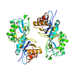
Crystal structure of endo-beta-N-acetylglucosaminidase H at high pH
Descriptor: Endo-beta-N-acetylglucosaminidase H, MAGNESIUM ION
Authors:Stachowski, T.R, Snell, M.E, Snell, E.S.
Deposit date:2019-12-28
Release date:2020-11-25
Last modified:2023-10-11
Method:X-RAY DIFFRACTION (2.1 Å)
Cite:SAXS studies of X-ray induced disulfide bond damage: Engineering high-resolution insight from a low-resolution technique.
Plos One, 15, 2020
8FUH
DownloadVisualize
BU of 8fuh by Molmil
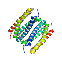
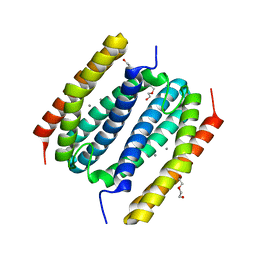
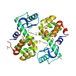
Rubrerythrin from B. pseudomallei: apo form
Descriptor: DI(HYDROXYETHYL)ETHER, Rubrerythrin
Authors:Monteiro, D.C.F, Snell, M.E, Budziszewski, G.R, Bowman, S.E.J.
Deposit date:2023-01-17
Release date:2024-01-31
Method:X-RAY DIFFRACTION (1.852 Å)
Cite:Rubrerythrin from B. pseudomallei: apo form
To Be Published
8FVV
DownloadVisualize
BU of 8fvv by Molmil
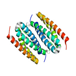
Rubrerythrin from B. pseudomallei: iron-bound
Descriptor: FE (III) ION, Rubrerythrin
Authors:Monteiro, D.C.F, Snell, M.E, Budziszewski, G.R, Bowman, S.E.J.
Deposit date:2023-01-19
Release date:2024-01-31
Method:X-RAY DIFFRACTION (1.93 Å)
Cite:Rubrerythrin from B. pseudomallei: iron-bound
To Be Published
8FXD
DownloadVisualize
BU of 8fxd by Molmil
Rubrerythrin from B. pseudomallei: manganese-bound
Descriptor: DI(HYDROXYETHYL)ETHER, MANGANESE (II) ION, Rubrerythrin
Authors:Monteiro, D.C.F, Snell, M.E, Budziszewski, G.R, Bowman, S.E.J.
Deposit date:2023-01-24
Release date:2024-02-07
Method:X-RAY DIFFRACTION (1.58 Å)
Cite:Rubrerythrin from B. pseudomallei: manganese-bound
To Be Published
6OBY
DownloadVisualize
BU of 6oby by Molmil
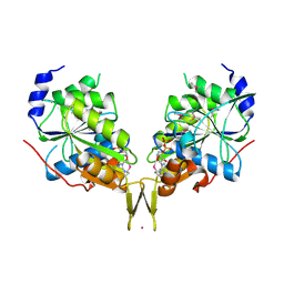
The nucleotide-binding protein AF_226 in complex with ADP from Archaeoglobus fulgidus with Co found by PIXE. Based on 3KB1.
Descriptor: ADENOSINE-5'-DIPHOSPHATE, COBALT (II) ION, Iron-sulfur cluster carrier protein
Authors:Snell, E.H, Garman, E.F, Lowe, E.D.
Deposit date:2019-03-21
Release date:2019-12-25
Last modified:2024-11-06
Method:X-RAY DIFFRACTION (2.87 Å)
Cite:High-Throughput PIXE as an Essential Quantitative Assay for Accurate Metalloprotein Structural Analysis: Development and Application.
J.Am.Chem.Soc., 142, 2020
6NLR
DownloadVisualize
BU of 6nlr by Molmil
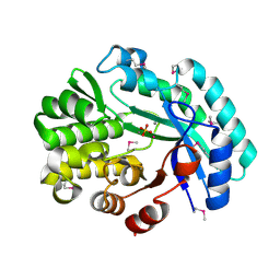
Crystal structure of the putative histidinol phosphatase hisK from Listeria monocytogenes with trinuclear metals determined by PIXE revealing sulphate ion in active site. Based on PIXE analysis and original date from 3DCP
Descriptor: CALCIUM ION, COBALT (II) ION, FE (III) ION, ...
Authors:Snell, E.H, Garman, E.F, Lowe, E.D.
Deposit date:2019-01-09
Release date:2019-12-25
Last modified:2024-10-23
Method:X-RAY DIFFRACTION (2.1 Å)
Cite:High-Throughput PIXE as an Essential Quantitative Assay for Accurate Metalloprotein Structural Analysis: Development and Application.
J.Am.Chem.Soc., 142, 2020
6OE2
DownloadVisualize
BU of 6oe2 by Molmil
X-Ray Structure of the C-terminal domain (277-440) of Putative chitobiase from Bacteroides thetaiotaomicron. Northeast Structural Genomics Consortium Target BtR324A. Re-refinement of 3GGL with correct metal Mn replacing Zn. New metal confirmed with PIXE analysis of original sample.
Descriptor: Chitobiase, DI(HYDROXYETHYL)ETHER, MANGANESE (II) ION
Authors:Snell, E.H, Garman, E.F, Lowe, E.D.
Deposit date:2019-03-27
Release date:2019-12-25
Last modified:2023-11-15
Method:X-RAY DIFFRACTION (3 Å)
Cite:High-Throughput PIXE as an Essential Quantitative Assay for Accurate Metalloprotein Structural Analysis: Development and Application.
J.Am.Chem.Soc., 142, 2020
3TL4
DownloadVisualize
BU of 3tl4 by Molmil
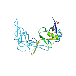
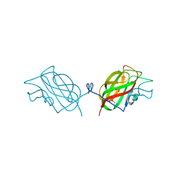
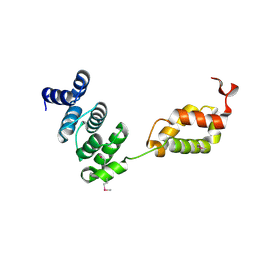
Crystal Structure of the tRNA Binding Domain of Glutaminyl-tRNA Synthetase from Saccharomyces cerevisiae
Descriptor: Glutaminyl-tRNA synthetase
Authors:Grant, T.D, Snell, E.H.
Deposit date:2011-08-29
Release date:2011-11-30
Last modified:2024-10-16
Method:X-RAY DIFFRACTION (2.3 Å)
Cite:Structural conservation of an ancient tRNA sensor in eukaryotic glutaminyl-tRNA synthetase.
Nucleic Acids Res., 40, 2012
8FW1
DownloadVisualize
BU of 8fw1 by Molmil
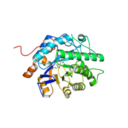
Gluconobacter Ene-Reductase (GluER) mutant - PagER
Descriptor: FLAVIN MONONUCLEOTIDE, N-ethylmaleimide reductase
Authors:Dahagam, S, Page, C, Patterson, M.G, Hyster, T.K.
Deposit date:2023-01-20
Release date:2023-06-28
Last modified:2023-10-25
Method:X-RAY DIFFRACTION (1.5 Å)
Cite:Regioselective Radical Alkylation of Arenes Using Evolved Photoenzymes.
J.Am.Chem.Soc., 145, 2023
4H3S
DownloadVisualize
BU of 4h3s by Molmil
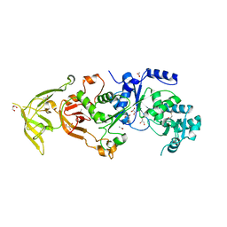
The Structure of Glutaminyl-tRNA Synthetase from Saccharomyces Cerevisiae
Descriptor: 2,3-DIHYDROXY-1,4-DITHIOBUTANE, ACETATE ION, BROMIDE ION, ...
Authors:Snell, E.H, Grant, T.D.
Deposit date:2012-09-14
Release date:2013-04-24
Last modified:2023-09-20
Method:X-RAY DIFFRACTION (2.15 Å)
Cite:The Structure of Yeast Glutaminyl-tRNA Synthetase and Modeling of Its Interaction with tRNA.
J.Mol.Biol., 425, 2013

227111

数据于2024-11-06公开中

PDB statisticsPDBj update infoContact PDBjnumon