Loading
PDBj
MenuPDBj@FacebookPDBj@TwitterPDBj@YouTubewwPDB FoundationwwPDB
RCSB PDBPDBeBMRBAdv. SearchSearch help
Search by PDB author
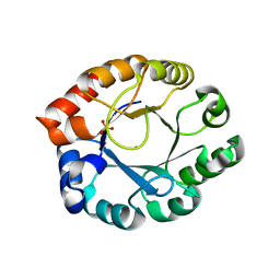
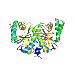
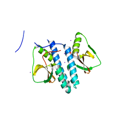
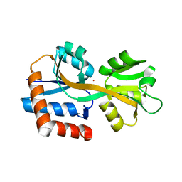
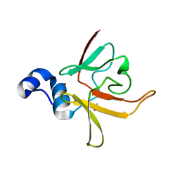
3CEZ
DownloadVisualize
BU of 3cez by Molmil
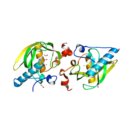
Crystal structure of methionine-R-sulfoxide reductase from Burkholderia pseudomallei
Descriptor: ACETIC ACID, Methionine-R-sulfoxide reductase, ZINC ION
Authors:Staker, B, Napuli, A, Nakazawa, S.H, Castaneda, L, Alkafeef, S, Vanvoorhis, W, Stewart, L, Myler, P, Seattle Structural Genomics Center for Infectious Disease (SSGCID)
Deposit date:2008-02-29
Release date:2008-03-18
Last modified:2023-08-30
Method:X-RAY DIFFRACTION (2.1 Å)
Cite:Methionine-R-sulfoxide reductase from Burkholderia pseudomallei.
To be Published
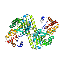
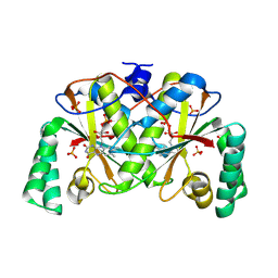
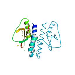
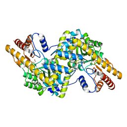
1TQX
DownloadVisualize
BU of 1tqx by Molmil
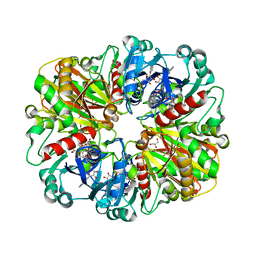
Crystal Structure of Pfal009167 A Putative D-Ribulose 5-Phosphate 3-Epimerase from P.falciparum
Descriptor: D-ribulose-5-phosphate 3-epimerase, putative, SULFATE ION, ...
Authors:Caruthers, J, Bosch, J, Hol, W.G.J, Structural Genomics of Pathogenic Protozoa Consortium (SGPP)
Deposit date:2004-06-18
Release date:2004-12-21
Last modified:2023-08-23
Method:X-RAY DIFFRACTION (2 Å)
Cite:Structure of a ribulose 5-phosphate 3-epimerase from Plasmodium falciparum.
Proteins, 62, 2006
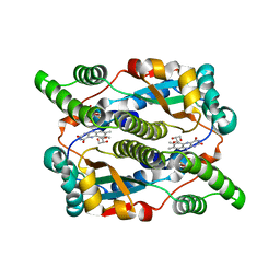
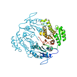
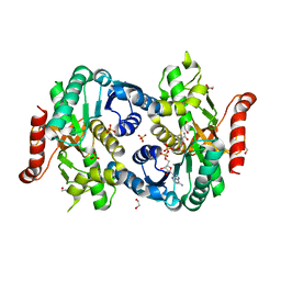
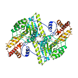
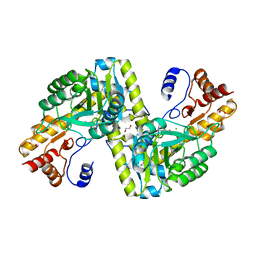
4W5K
DownloadVisualize
BU of 4w5k by Molmil
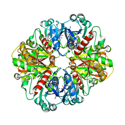
Structure of a mitochondrial aspartate aminotransferase from Trypanosoma brucei, K237A mutant
Descriptor: 1,2-ETHANEDIOL, Aspartate aminotransferase, mitochondrial, ...
Authors:Seattle Structural Genomics Center for Infectious Disease (SSGCID)
Deposit date:2014-08-18
Release date:2014-09-24
Last modified:2023-09-27
Method:X-RAY DIFFRACTION (1.7 Å)
Cite:Structures of aspartate aminotransferases from Trypanosoma brucei, Leishmania major and Giardia lamblia.
Acta Crystallogr.,Sect.F, 71, 2015
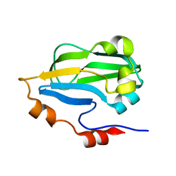
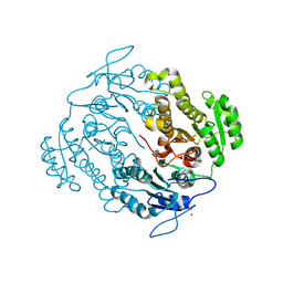
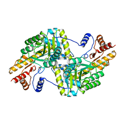
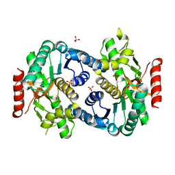
8DOR
DownloadVisualize
BU of 8dor by Molmil
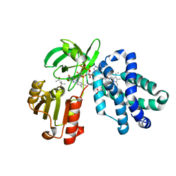
Crystal structure of Dihydropteridine reductase/oxygen-insensitive NAD(P)H nitroreductase from Klebsiella pneumoniae
Descriptor: Dihydropteridine reductase/oxygen-insensitive NAD(P)H nitroreductase, FLAVIN MONONUCLEOTIDE, GLYCEROL, ...
Authors:Seattle Structural Genomics Center for Infectious Disease (SSGCID)
Deposit date:2022-07-14
Release date:2022-07-27
Last modified:2024-08-21
Method:X-RAY DIFFRACTION (1.35 Å)
Cite:Crystal structures of NAD(P)H nitroreductases from Klebsiella pneumoniae.
Acta Crystallogr.,Sect.F, 80, 2024
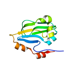
3HGB
DownloadVisualize
BU of 3hgb by Molmil
Crystal structure of glycine cleavage system protein H from Mycobacterium tuberculosis
Descriptor: Glycine cleavage system H protein
Authors:Seattle Structural Genomics Center for Infectious Disease (SSGCID)
Deposit date:2009-05-13
Release date:2009-05-26
Last modified:2023-09-06
Method:X-RAY DIFFRACTION (1.75 Å)
Cite:X-ray structure determination of the glycine cleavage system protein H of Mycobacterium tuberculosis using an inverse Compton synchrotron X-ray source.
J.Struct.Funct.Genom., 11, 2010
7TMF
DownloadVisualize
BU of 7tmf by Molmil
Crystal Structure of NAD(P)H nitroreductase from Klebsiella pneumoniae (short b-axis)
Descriptor: 2-[BIS-(2-HYDROXY-ETHYL)-AMINO]-2-HYDROXYMETHYL-PROPANE-1,3-DIOL, FLAVIN MONONUCLEOTIDE, GLYCEROL, ...
Authors:Seattle Structural Genomics Center for Infectious Disease (SSGCID)
Deposit date:2022-01-19
Release date:2022-02-02
Last modified:2024-08-14
Method:X-RAY DIFFRACTION (1.97 Å)
Cite:Crystal structures of NAD(P)H nitroreductases from Klebsiella pneumoniae
Acta Crystallogr.,Sect.F, F80, 2024
7TMG
DownloadVisualize
BU of 7tmg by Molmil
Crystal Structure of NAD(P)H nitroreductase from Klebsiella pneumoniae (long b-axis)
Descriptor: FLAVIN MONONUCLEOTIDE, NAD(P)H nitroreductase, PHOSPHATE ION
Authors:Seattle Structural Genomics Center for Infectious Disease (SSGCID)
Deposit date:2022-01-19
Release date:2022-02-02
Last modified:2024-08-14
Method:X-RAY DIFFRACTION (1.9 Å)
Cite:Crystal structures of NAD(P)H nitroreductases from Klebsiella pneumoniae
Acta Crystallogr.,Sect.F, F80, 2024
5VP5
DownloadVisualize
BU of 5vp5 by Molmil
Crystal structure of a 3-oxoacyl-acyl-carrier protein reductase FabG4 from Mycobacterium smegmatis bound to NAD
Descriptor: 3-oxoacyl-acyl-carrier protein reductase FabG4, CHLORIDE ION, NICOTINAMIDE-ADENINE-DINUCLEOTIDE
Authors:Seattle Structural Genomics Center for Infectious Disease (SSGCID)
Deposit date:2017-05-04
Release date:2017-05-17
Last modified:2024-07-17
Method:X-RAY DIFFRACTION (1.8 Å)
Cite:Structural and functional characterization of FabG4 from Mycolicibacterium smegmatis.
Acta Crystallogr.,Sect.F, 80, 2024
3U0B
DownloadVisualize
BU of 3u0b by Molmil
Crystal structure of an oxidoreductase from Mycobacterium smegmatis
Descriptor: Oxidoreductase, short chain dehydrogenase/reductase family protein, SODIUM ION
Authors:Arakaki, T.L, Staker, B.L, Clifton, M.C, Abendroth, J, Seattle Structural Genomics Center for Infectious Disease (SSGCID)
Deposit date:2011-09-28
Release date:2011-10-05
Last modified:2024-07-17
Method:X-RAY DIFFRACTION (1.7 Å)
Cite:Structural and functional characterization of FabG4 from Mycolicibacterium smegmatis.
Acta Crystallogr.,Sect.F, 80, 2024
5KOB
DownloadVisualize
BU of 5kob by Molmil
Crystal structure of a peptide deformylase from Burkholderia xenovorans
Descriptor: 1,2-ETHANEDIOL, FE (II) ION, FORMIC ACID, ...
Authors:SSGCID, Seattle Structural Genomics Center for Infectious Disease (SSGCID)
Deposit date:2016-06-30
Release date:2016-07-13
Last modified:2023-10-04
Method:X-RAY DIFFRACTION (1.6 Å)
Cite:Crystal structure of a peptide deformylase from Burkholderia xenovorans
to be published
2B4R
DownloadVisualize
BU of 2b4r by Molmil
Crystal structure of glyceraldehyde-3-phosphate dehydrogenase from Plasmodium falciparum at 2.25 Angstrom Resolution reveals intriguing extra electron density in the active site
Descriptor: 4-(2-AMINOETHYL)BENZENESULFONYL FLUORIDE, GLYCEROL, NICOTINAMIDE-ADENINE-DINUCLEOTIDE, ...
Authors:Robien, M.A, Bosch, J, Hol, W.G.J, Structural Genomics of Pathogenic Protozoa Consortium (SGPP)
Deposit date:2005-09-26
Release date:2005-10-04
Last modified:2023-08-23
Method:X-RAY DIFFRACTION (2.25 Å)
Cite:Crystal structure of glyceraldehyde-3-phosphate dehydrogenase from Plasmodium falciparum at 2.25 A resolution reveals intriguing extra electron density in the active site
Proteins, 62, 2006
2B4T
DownloadVisualize
BU of 2b4t by Molmil
Crystal structure of glyceraldehyde-3-phosphate dehydrogenase from Plasmodium falciparum at 2.25 Angstrom resolution reveals intriguing extra electron density in the active site
Descriptor: 4-(2-AMINOETHYL)BENZENESULFONYL FLUORIDE, NICOTINAMIDE-ADENINE-DINUCLEOTIDE, glyceraldehyde-3-phosphate dehydrogenase
Authors:Robien, M.A, Bosch, J, Hol, W.G.J, Structural Genomics of Pathogenic Protozoa Consortium (SGPP)
Deposit date:2005-09-26
Release date:2005-10-04
Last modified:2023-08-23
Method:X-RAY DIFFRACTION (2.5 Å)
Cite:Crystal structure of glyceraldehyde-3-phosphate dehydrogenase from Plasmodium falciparum at 2.25 A resolution reveals intriguing extra electron density in the active site
Proteins, 62, 2006
6O0A
DownloadVisualize
BU of 6o0a by Molmil
Crystal structure of flavohemoglobin from Malassezia yamatoensis with bound FAD and heme determined by iron SAD phasing
Descriptor: (4S)-2-METHYL-2,4-PENTANEDIOL, FLAVIN-ADENINE DINUCLEOTIDE, Flavohemoglobin, ...
Authors:Seattle Structural Genomics Center for Infectious Disease (SSGCID)
Deposit date:2019-02-15
Release date:2019-03-06
Last modified:2024-05-22
Method:X-RAY DIFFRACTION (1.7 Å)
Cite:HGT in the human and skin commensal Malassezia : A bacterially derived flavohemoglobin is required for NO resistance and host interaction.
Proc.Natl.Acad.Sci.USA, 117, 2020
4I1I
DownloadVisualize
BU of 4i1i by Molmil
Crystal structure of a putative Cytosolic malate dehydrogenase from Leishmania major Friedlin in complex with NAD
Descriptor: 1,2-ETHANEDIOL, Malate dehydrogenase, NICOTINAMIDE-ADENINE-DINUCLEOTIDE, ...
Authors:Seattle Structural Genomics Center for Infectious Disease, Seattle Structural Genomics Center for Infectious Disease (SSGCID)
Deposit date:2012-11-21
Release date:2012-12-19
Last modified:2023-09-20
Method:X-RAY DIFFRACTION (1.5 Å)
Cite:Crystal structure of a putative Cytosolic malate dehydrogenase from Leishmania major Friedlin in complex with NAD
TO BE PUBLISHED
3IFT
DownloadVisualize
BU of 3ift by Molmil
Crystal structure of glycine cleavage system protein H from Mycobacterium tuberculosis, using X-rays from the Compact Light Source.
Descriptor: Glycine cleavage system H protein
Authors:Edwards, T.E, Abendroth, J, Staker, B, Mayer, C, Phan, I, Kelley, A, Analau, E, Leibly, D, Rifkin, J, Loewen, R, Ruth, R.D, Stewart, L.J, Accelerated Technologies Center for Gene to 3D Structure (ATCG3D)
Deposit date:2009-07-25
Release date:2009-08-11
Last modified:2023-09-06
Method:X-RAY DIFFRACTION (2 Å)
Cite:X-ray structure determination of the glycine cleavage system protein H of Mycobacterium tuberculosis using an inverse Compton synchrotron X-ray source.
J.Struct.Funct.Genom., 11, 2010
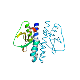
3LR3
DownloadVisualize
BU of 3lr3 by Molmil
Periplasmic domain of the risS sensor protein from Burkholderia pesuromallei, low pH native structure
Descriptor: Sensor protein
Authors:Seattle Structural Genomics Center for Infectious Disease (SSGCID)
Deposit date:2010-02-10
Release date:2010-06-16
Last modified:2023-09-06
Method:X-RAY DIFFRACTION (2.1 Å)
Cite:Structural basis for pH sensing by the periplasmic domain of the risS histidine kinase from Burkholderia pseudomallei
To be Published
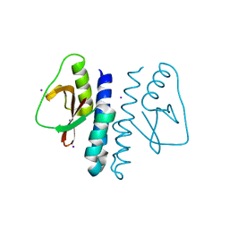
3LR0
DownloadVisualize
BU of 3lr0 by Molmil
Periplasmic domain of the risS sensor protein from Burkholderia pseudomallei, iodide phased at low pH
Descriptor: IODIDE ION, Sensor protein
Authors:Seattle Structural Genomics Center for Infectious Disease (SSGCID)
Deposit date:2010-02-10
Release date:2010-06-16
Last modified:2024-02-21
Method:X-RAY DIFFRACTION (1.9 Å)
Cite:Structural basis for pH sensing by the periplasmic domain of the risS histidine kinase from Bukholderia pseudomallei
To be Published
3LR5
DownloadVisualize
BU of 3lr5 by Molmil
Periplasmic domain of the risS sensor protein from Burkholderia pseudomallei, iodide phased at neutral pH
Descriptor: IODIDE ION, Sensor protein
Authors:Seattle Structural Genomics Center for Infectious Disease (SSGCID)
Deposit date:2010-02-10
Release date:2010-06-16
Last modified:2024-02-21
Method:X-RAY DIFFRACTION (2.3 Å)
Cite:Structural basis for pH sensing by the periplsamic domain of the risS histidine kinase from Burkholderia pseudomallei
To be Published
3LR4
DownloadVisualize
BU of 3lr4 by Molmil
Periplasmic domain of the risS sensor protein from Burkholderia pseudomallei, barium phased at low pH
Descriptor: BARIUM ION, CHLORIDE ION, Sensor protein
Authors:Seattle Structural Genomics Center for Infectious Disease (SSGCID)
Deposit date:2010-02-10
Release date:2010-06-16
Last modified:2024-02-21
Method:X-RAY DIFFRACTION (1.9 Å)
Cite:Structural basis for pH sensing by the periplasmic domain of the risS histidine kinase from Burkholderia pseudomallei
To be Published
3MEB
DownloadVisualize
BU of 3meb by Molmil
Structure of cytoplasmic aspartate aminotransferase from giardia lamblia
Descriptor: 1,2-ETHANEDIOL, Aspartate aminotransferase, PYRIDOXAL-5'-PHOSPHATE, ...
Authors:Seattle Structural Genomics Center for Infectious Disease (SSGCID)
Deposit date:2010-03-31
Release date:2010-04-21
Last modified:2023-09-06
Method:X-RAY DIFFRACTION (1.9 Å)
Cite:Structures of aspartate aminotransferases from Trypanosoma brucei, Leishmania major and Giardia lamblia.
Acta Crystallogr F Struct Biol Commun, 71, 2015
4F4E
DownloadVisualize
BU of 4f4e by Molmil
Crystal structure of Aromatic-amino-acid aminotransferase from Burkholderia pseudomallei covalently bound to pyridoxal phosphate
Descriptor: 1,2-ETHANEDIOL, Aromatic-amino-acid aminotransferase
Authors:Seattle Structural Genomics Center for Infectious Disease (SSGCID)
Deposit date:2012-05-10
Release date:2012-05-23
Last modified:2023-12-06
Method:X-RAY DIFFRACTION (1.8 Å)
Cite:Crystal structure of Aromatic-amino-acid aminotransferase from Burkholderia pseudomallei covalently bound to pyridoxal phosphate
To be Published
4F3P
DownloadVisualize
BU of 4f3p by Molmil
Crystal structure of a Glutamine-binding periplasmic protein from Burkholderia pseudomallei in complex with glutamine
Descriptor: Glutamine-binding periplasmic protein
Authors:Seattle Structural Genomics Center for Infectious Disease (SSGCID)
Deposit date:2012-05-09
Release date:2012-05-23
Last modified:2023-09-13
Method:X-RAY DIFFRACTION (2.4 Å)
Cite:Crystal structure of a Glutamine-binding periplasmic protein from Burkholderia pseudomallei in complex with glutamine
To be Published
4H51
DownloadVisualize
BU of 4h51 by Molmil
Crystal structure of a putative Aspartate Aminotransferase from Leishmania major Friedlin
Descriptor: 1,2-ETHANEDIOL, Aspartate aminotransferase
Authors:Seattle Structural Genomics Center for Infectious Disease (SSGCID)
Deposit date:2012-09-18
Release date:2012-10-10
Last modified:2015-05-20
Method:X-RAY DIFFRACTION (1.85 Å)
Cite:Structures of aspartate aminotransferases from Trypanosoma brucei, Leishmania major and Giardia lamblia.
Acta Crystallogr F Struct Biol Commun, 71, 2015
4EU1
DownloadVisualize
BU of 4eu1 by Molmil
Structure of a mitochondrial aspartate aminotransferase from Trypanosoma brucei
Descriptor: 1,2-ETHANEDIOL, CHLORIDE ION, Mitochondrial aspartate aminotransferase
Authors:Seattle Structural Genomics Center for Infectious Disease (SSGCID)
Deposit date:2012-04-25
Release date:2012-05-16
Last modified:2023-12-06
Method:X-RAY DIFFRACTION (2.3 Å)
Cite:Structures of aspartate aminotransferases from Trypanosoma brucei, Leishmania major and Giardia lamblia.
Acta Crystallogr F Struct Biol Commun, 71, 2015
4H7P
DownloadVisualize
BU of 4h7p by Molmil
Crystal structure of a putative Cytosolic malate dehydrogenase from Leishmania major Friedlin
Descriptor: Malate dehydrogenase, SULFATE ION
Authors:Seattle Structural Genomics Center for Infectious Disease (SSGCID)
Deposit date:2012-09-20
Release date:2012-10-10
Last modified:2023-09-20
Method:X-RAY DIFFRACTION (1.3 Å)
Cite:Crystal structure of a putative Cytosolic malate dehydrogenase from Leishmania major Friedlin
To be Published


12>

227111

数据于2024-11-06公开中

PDB statisticsPDBj update infoContact PDBjnumon