Loading
PDBj
MenuPDBj@FacebookPDBj@TwitterPDBj@YouTubewwPDB FoundationwwPDB
RCSB PDBPDBeBMRBAdv. SearchSearch help
Search by PDB author
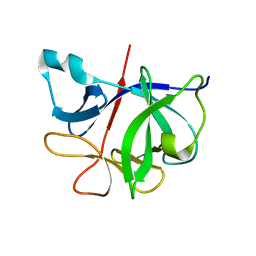
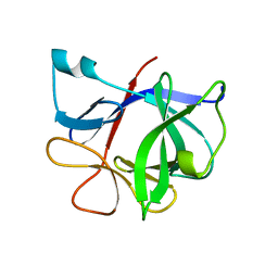
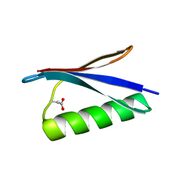
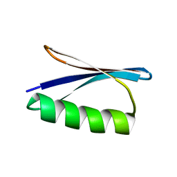
1GB1
DownloadVisualize
BU of 1gb1 by Molmil
A NOVEL, HIGHLY STABLE FOLD OF THE IMMUNOGLOBULIN BINDING DOMAIN OF STREPTOCOCCAL PROTEIN G
Descriptor: PROTEIN G
Authors:Gronenborn, A.M, Clore, G.M.
Deposit date:1991-05-15
Release date:1993-04-15
Last modified:2024-05-22
Method:SOLUTION NMR
Cite:A novel, highly stable fold of the immunoglobulin binding domain of streptococcal protein G.
Science, 253, 1991
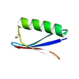
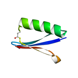
2GB1
DownloadVisualize
BU of 2gb1 by Molmil
A NOVEL, HIGHLY STABLE FOLD OF THE IMMUNOGLOBULIN BINDING DOMAIN OF STREPTOCOCCAL PROTEIN G
Descriptor: PROTEIN G
Authors:Gronenborn, A.M, Clore, G.M.
Deposit date:1991-05-15
Release date:1993-04-15
Last modified:2024-05-29
Method:SOLUTION NMR
Cite:A novel, highly stable fold of the immunoglobulin binding domain of streptococcal protein G.
Science, 253, 1991
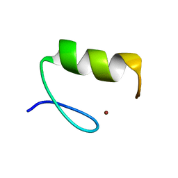
3ZNF
DownloadVisualize
BU of 3znf by Molmil
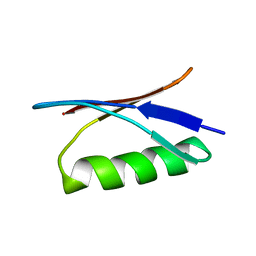
HIGH-RESOLUTION THREE-DIMENSIONAL STRUCTURE OF A SINGLE ZINC FINGER FROM A HUMAN ENHANCER BINDING PROTEIN IN SOLUTION
Descriptor: ZINC FINGER, ZINC ION
Authors:Gronenborn, A.M, Clore, G.M, Omichinski, J.G.
Deposit date:1990-07-09
Release date:1992-01-15
Last modified:2024-05-22
Method:SOLUTION NMR
Cite:High-resolution three-dimensional structure of a single zinc finger from a human enhancer binding protein in solution.
Biochemistry, 29, 1990
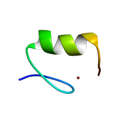
4ZNF
DownloadVisualize
BU of 4znf by Molmil
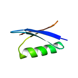
HIGH-RESOLUTION THREE-DIMENSIONAL STRUCTURE OF A SINGLE ZINC FINGER FROM A HUMAN ENHANCER BINDING PROTEIN IN SOLUTION
Descriptor: ZINC FINGER, ZINC ION
Authors:Gronenborn, A.M, Clore, G.M, Omichinski, J.G.
Deposit date:1990-07-09
Release date:1992-01-15
Last modified:2024-05-22
Method:SOLUTION NMR
Cite:High-resolution three-dimensional structure of a single zinc finger from a human enhancer binding protein in solution.
Biochemistry, 29, 1990
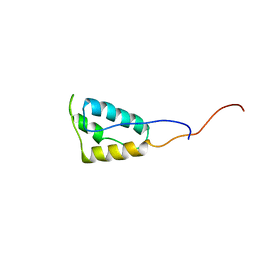
5T82
DownloadVisualize
BU of 5t82 by Molmil
HIV-1 reverse transcriptase thumb subdomain
Descriptor: Reverse transcriptase
Authors:Gronenborn, A.M, Sharaf, N.G, Byeon, I.-J.L.
Deposit date:2016-09-06
Release date:2017-01-18
Last modified:2024-05-15
Method:SOLUTION NMR
Cite:NMR structure of the HIV-1 reverse transcriptase thumb subdomain.
J. Biomol. NMR, 66, 2016
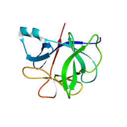
6I1B
DownloadVisualize
BU of 6i1b by Molmil
HIGH-RESOLUTION THREE-DIMENSIONAL STRUCTURE OF INTERLEUKIN-1 BETA IN SOLUTION BY THREE-AND FOUR-DIMENSIONAL NUCLEAR MAGNETIC RESONANCE SPECTROSCOPY
Descriptor: INTERLEUKIN-1 BETA
Authors:Clore, G.M, Gronenborn, A.M.
Deposit date:1991-01-22
Release date:1992-10-15
Last modified:2024-05-22
Method:SOLUTION NMR
Cite:High-resolution three-dimensional structure of interleukin 1 beta in solution by three- and four-dimensional nuclear magnetic resonance spectroscopy.
Biochemistry, 30, 1991
9ILB
DownloadVisualize
BU of 9ilb by Molmil
HUMAN INTERLEUKIN-1 BETA
Descriptor: PROTEIN (HUMAN INTERLEUKIN-1 BETA)
Authors:Yu, B, Blaber, M, Gronenborn, A.M, Clore, G.M, Caspar, D.L.D.
Deposit date:1998-10-22
Release date:1999-01-06
Last modified:2023-09-20
Method:X-RAY DIFFRACTION (2.28 Å)
Cite:Disordered water within a hydrophobic protein cavity visualized by x-ray crystallography.
Proc.Natl.Acad.Sci.USA, 96, 1999
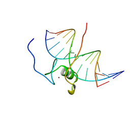
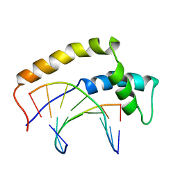
7GAT
DownloadVisualize
BU of 7gat by Molmil
SOLUTION NMR STRUCTURE OF THE L22V MUTANT DNA BINDING DOMAIN OF AREA COMPLEXED TO A 13 BP DNA CONTAINING A TGATA SITE, 34 STRUCTURES
Descriptor: DNA (5'-D(*CP*AP*GP*TP*GP*AP*TP*AP*GP*AP*GP*AP*C)-3'), DNA (5'-D(*GP*TP*CP*TP*CP*TP*AP*TP*CP*AP*CP*TP*G)-3'), NITROGEN REGULATORY PROTEIN AREA, ...
Authors:Clore, G.M, Starich, M, Wikstrom, M, Gronenborn, A.M.
Deposit date:1997-11-07
Release date:1998-01-28
Last modified:2024-05-22
Method:SOLUTION NMR
Cite:The solution structure of the Leu22-->Val mutant AREA DNA binding domain complexed with a TGATAG core element defines a role for hydrophobic packing in the determination of specificity.
J.Mol.Biol., 277, 1998
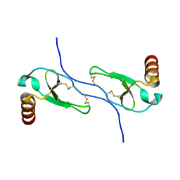
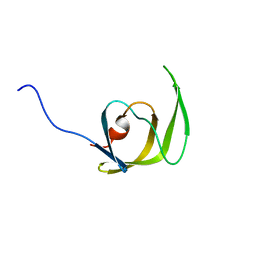
1HUN
DownloadVisualize
BU of 1hun by Molmil
SOLUTION STRUCTURE OF THE CHEMOKINE HMIP-1BETA(SLASH)ACT-2 BY MULTI-DIMENSIONAL NMR: A NOVEL CHEMOKINE DIMER
Descriptor: HUMAN MACROPHAGE INFLAMMATORY PROTEIN 1 BETA
Authors:Clore, G.M, Lodi, P.J, Garrett, D.S, Gronenborn, A.M.
Deposit date:1994-01-31
Release date:1994-04-30
Last modified:2022-02-23
Method:SOLUTION NMR
Cite:High-resolution solution structure of the beta chemokine hMIP-1 beta by multidimensional NMR.
Science, 263, 1994
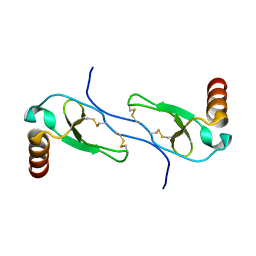
1HUM
DownloadVisualize
BU of 1hum by Molmil
SOLUTION STRUCTURE OF THE CHEMOKINE HMIP-1BETA(SLASH)ACT-2 BY MULTI-DIMENSIONAL NMR: A NOVEL CHEMOKINE DIMER
Descriptor: HUMAN MACROPHAGE INFLAMMATORY PROTEIN 1 BETA
Authors:Clore, G.M, Lodi, P.J, Garrett, D.S, Gronenborn, A.M.
Deposit date:1994-01-31
Release date:1994-04-30
Last modified:2024-10-23
Method:SOLUTION NMR
Cite:High-resolution solution structure of the beta chemokine hMIP-1 beta by multidimensional NMR.
Science, 263, 1994
7I1B
DownloadVisualize
BU of 7i1b by Molmil
HIGH-RESOLUTION THREE-DIMENSIONAL STRUCTURE OF INTERLEUKIN-1 BETA IN SOLUTION BY THREE-AND FOUR-DIMENSIONAL NUCLEAR MAGNETIC RESONANCE SPECTROSCOPY
Descriptor: INTERLEUKIN-1 BETA
Authors:Clore, G.M, Gronenborn, A.M.
Deposit date:1991-01-22
Release date:1992-10-15
Last modified:2024-05-22
Method:SOLUTION NMR
Cite:High-resolution three-dimensional structure of interleukin 1 beta in solution by three- and four-dimensional nuclear magnetic resonance spectroscopy.
Biochemistry, 30, 1991
1HRZ
DownloadVisualize
BU of 1hrz by Molmil
THE 3D STRUCTURE OF THE HUMAN SRY-DNA COMPLEX SOLVED BY MULTI-DIMENSIONAL HETERONUCLEAR-EDITED AND-FILTERED NMR
Descriptor: DNA (5'-D(*GP*CP*AP*CP*AP*AP*AP*C)-3'), DNA (5'-D(*GP*TP*TP*TP*GP*TP*GP*C)-3'), HUMAN SRY
Authors:Clore, G.M, Werner, M.H, Huth, J.R, Gronenborn, A.M.
Deposit date:1995-05-09
Release date:1995-09-15
Last modified:2024-05-22
Method:SOLUTION NMR
Cite:Molecular basis of human 46X,Y sex reversal revealed from the three-dimensional solution structure of the human SRY-DNA complex.
Cell(Cambridge,Mass.), 81, 1995
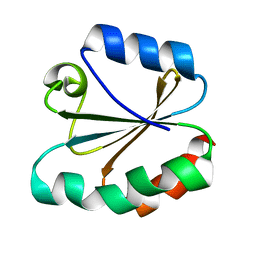
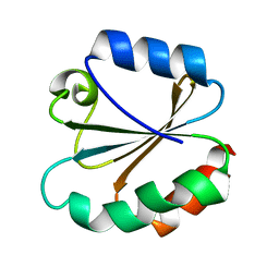
4TRX
DownloadVisualize
BU of 4trx by Molmil
HIGH-RESOLUTION THREE-DIMENSIONAL STRUCTURE OF REDUCED RECOMBINANT HUMAN THIOREDOXIN IN SOLUTION
Descriptor: THIOREDOXIN
Authors:Forman-Kay, J.D, Clore, G.M, Gronenborn, A.M.
Deposit date:1990-12-17
Release date:1992-01-15
Last modified:2024-05-22
Method:SOLUTION NMR
Cite:High-resolution three-dimensional structure of reduced recombinant human thioredoxin in solution.
Biochemistry, 30, 1991
1Q9P
DownloadVisualize
BU of 1q9p by Molmil
Solution structure of the mature HIV-1 protease monomer
Descriptor: HIV-1 Protease
Authors:Ishima, R, Torchia, D.A, Lynch, S.M, Gronenborn, A.M, Louis, J.M.
Deposit date:2003-08-25
Release date:2004-03-02
Last modified:2024-05-22
Method:SOLUTION NMR
Cite:Solution structure of the mature HIV-1 protease monomer: Insight into the tertiary fold and stability of a precursor
J.Biol.Chem., 278, 2003
1QWV
DownloadVisualize
BU of 1qwv by Molmil
Solution structure of Antheraea polyphemus pheromone binding protein (ApolPBP)
Descriptor: Pheromone-binding protein
Authors:Mohanty, S, Zubkov, S, Gronenborn, A.M.
Deposit date:2003-09-03
Release date:2004-03-23
Last modified:2024-10-30
Method:SOLUTION NMR
Cite:The Solution NMR Structure of Antheraea polyphemus PBP Provides New Insight into Pheromone Recognition by Pheromone-binding Proteins
J.Mol.Biol., 337, 2004
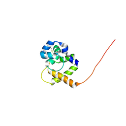
3TRX
DownloadVisualize
BU of 3trx by Molmil
HIGH-RESOLUTION THREE-DIMENSIONAL STRUCTURE OF REDUCED RECOMBINANT HUMAN THIOREDOXIN IN SOLUTION
Descriptor: THIOREDOXIN
Authors:Forman-Kay, J.D, Clore, G.M, Gronenborn, A.M.
Deposit date:1990-12-17
Release date:1992-01-15
Last modified:2024-05-22
Method:SOLUTION NMR
Cite:High-resolution three-dimensional structure of reduced recombinant human thioredoxin in solution.
Biochemistry, 30, 1991
8UM9
DownloadVisualize
BU of 8um9 by Molmil
Site-specific Aspartic Acid Dehydration and Isomerization in Streptococcal Protein GB1: D-Asp40 Variant
Descriptor: Immunoglobulin G-binding protein G
Authors:Heath, S.L, Guseman, A.J, Gronenborn, A.M, Horne, W.S.
Deposit date:2023-10-17
Release date:2024-05-01
Method:SOLUTION NMR
Cite:Probing effects of site-specific aspartic acid isomerization on structure and stability of GB1 through chemical protein synthesis.
Protein Sci., 33, 2024
8UMB
DownloadVisualize
BU of 8umb by Molmil
Site-specific Aspartic Acid Dehydration and Isomerization in Streptococcal Protein GB1: L-Aspartyl Succinimide 40-41 Variant
Descriptor: Immunoglobulin G-binding protein G
Authors:Heath, S.L, Guseman, A.J, Gronenborn, A.M, Horne, W.S.
Deposit date:2023-10-17
Release date:2024-05-01
Last modified:2024-10-09
Method:SOLUTION NMR
Cite:Probing effects of site-specific aspartic acid isomerization on structure and stability of GB1 through chemical protein synthesis.
Protein Sci., 33, 2024
8UM7
DownloadVisualize
BU of 8um7 by Molmil
Site-specific Aspartic Acid Dehydration and Isomerization in Streptococcal Protein GB1: Wild-type Protein
Descriptor: Immunoglobulin G-binding protein G
Authors:Heath, S.L, Guseman, A.J, Gronenborn, A.M, Horne, W.S.
Deposit date:2023-10-17
Release date:2024-05-01
Method:SOLUTION NMR
Cite:Probing effects of site-specific aspartic acid isomerization on structure and stability of GB1 through chemical protein synthesis.
Protein Sci., 33, 2024
8UMS
DownloadVisualize
BU of 8ums by Molmil
Site-specific Aspartic Acid Dehydration and Isomerization in Streptococcal Protein GB1: L-isoAsp40 Variant
Descriptor: Immunoglobulin G-binding protein G
Authors:Heath, S.L, Guseman, A.J, Gronenborn, A.M, Horne, W.S.
Deposit date:2023-10-18
Release date:2024-05-01
Last modified:2024-10-23
Method:SOLUTION NMR
Cite:Probing effects of site-specific aspartic acid isomerization on structure and stability of GB1 through chemical protein synthesis.
Protein Sci., 33, 2024
8UMA
DownloadVisualize
BU of 8uma by Molmil
Site-specific Aspartic Acid Dehydration and Isomerization in Streptococcal Protein GB1: D-isoAsp40 Variant
Descriptor: Immunoglobulin G-binding protein G
Authors:Heath, S.L, Guseman, A.J, Gronenborn, A.M, Horne, W.S.
Deposit date:2023-10-17
Release date:2024-05-01
Method:SOLUTION NMR
Cite:Probing effects of site-specific aspartic acid isomerization on structure and stability of GB1 through chemical protein synthesis.
Protein Sci., 33, 2024
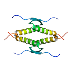
1SAK
DownloadVisualize
BU of 1sak by Molmil
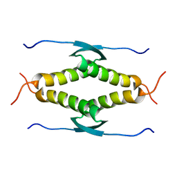
HIGH RESOLUTION SOLUTION NMR STRUCTURE OF THE OLIGOMERIZATION DOMAIN OF P53 BY MULTI-DIMENSIONAL NMR (SAC STRUCTURES)
Descriptor: TUMOR SUPPRESSOR P53
Authors:Clore, G.M, Omichinski, J.G, Gronenborn, A.M.
Deposit date:1995-03-12
Release date:1995-10-15
Last modified:2024-05-22
Method:SOLUTION NMR
Cite:Refined solution structure of the oligomerization domain of the tumour suppressor p53.
Nat.Struct.Biol., 2, 1995
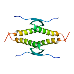
1SAE
DownloadVisualize
BU of 1sae by Molmil
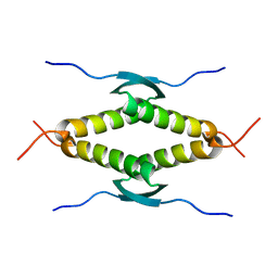
HIGH RESOLUTION SOLUTION NMR STRUCTURE OF THE OLIGOMERIZATION DOMAIN OF P53 BY MULTI-DIMENSIONAL NMR (SAC STRUCTURES)
Descriptor: TUMOR SUPPRESSOR P53
Authors:Clore, G.M, Omichinski, J.G, Gronenborn, A.M.
Deposit date:1995-03-12
Release date:1995-10-15
Last modified:2024-05-22
Method:SOLUTION NMR
Cite:Refined solution structure of the oligomerization domain of the tumour suppressor p53.
Nat.Struct.Biol., 2, 1995
1SAF
DownloadVisualize
BU of 1saf by Molmil
HIGH RESOLUTION SOLUTION NMR STRUCTURE OF THE OLIGOMERIZATION DOMAIN OF P53 BY MULTI-DIMENSIONAL NMR (SAD STRUCTURES)
Descriptor: TUMOR SUPPRESSOR P53
Authors:Clore, G.M, Omichinski, J.G, Gronenborn, A.M.
Deposit date:1995-03-12
Release date:1995-10-15
Last modified:2024-05-01
Method:SOLUTION NMR
Cite:Refined solution structure of the oligomerization domain of the tumour suppressor p53.
Nat.Struct.Biol., 2, 1995
1SAL
DownloadVisualize
BU of 1sal by Molmil
HIGH RESOLUTION SOLUTION NMR STRUCTURE OF THE OLIGOMERIZATION DOMAIN OF P53 BY MULTI-DIMENSIONAL NMR (SAD STRUCTURES)
Descriptor: TUMOR SUPPRESSOR P53
Authors:Clore, G.M, Omichinski, J.G, Gronenborn, A.M.
Deposit date:1995-03-12
Release date:1995-10-15
Last modified:2024-05-22
Method:SOLUTION NMR
Cite:Refined solution structure of the oligomerization domain of the tumour suppressor p53.
Nat.Struct.Biol., 2, 1995

226707

数据于2024-10-30公开中

PDB statisticsPDBj update infoContact PDBjnumon