Loading
PDBj
MenuPDBj@FacebookPDBj@X(formerly Twitter)PDBj@BlueSkyPDBj@YouTubewwPDB FoundationwwPDB
RCSB PDBPDBeBMRBAdv. SearchSearch help
Search by PDB author
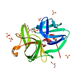
2H5D
DownloadVisualize
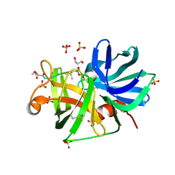
BU of 2h5d by Molmil
0.9A resolution crystal structure of alpha-lytic protease complexed with a transition state analogue, MeOSuc-Ala-Ala-Pro-Val boronic acid
Descriptor: ALPHA-LYTIC PROTEASE, GLYCEROL, MEOSUC-ALA-ALA-PRO-ALA BORONIC ACID INHIBITOR, ...
Authors:Fuhrmann, C.N, Agard, D.A.
Deposit date:2006-05-25
Release date:2006-09-26
Last modified:2023-11-15
Method:X-RAY DIFFRACTION (0.9 Å)
Cite:Subangstrom crystallography reveals that short ionic hydrogen bonds, and not a His-Asp low-barrier hydrogen bond, stabilize the transition state in serine protease catalysis
J.Am.Chem.Soc., 128, 2006
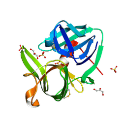
2H5C
DownloadVisualize
BU of 2h5c by Molmil
0.82A resolution crystal structure of alpha-lytic protease at pH 5
Descriptor: ALPHA-LYTIC PROTEASE, GLYCEROL, SULFATE ION
Authors:Fuhrmann, C.N, Daugherty, M.D, Agard, D.A.
Deposit date:2006-05-25
Release date:2006-09-26
Last modified:2024-11-06
Method:X-RAY DIFFRACTION (0.82 Å)
Cite:Subangstrom crystallography reveals that short ionic hydrogen bonds, and not a His-Asp low-barrier hydrogen bond, stabilize the transition state in serine protease catalysis
J.Am.Chem.Soc., 128, 2006
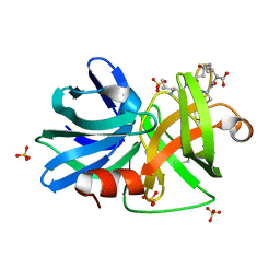
1SSX
DownloadVisualize
BU of 1ssx by Molmil
0.83A resolution crystal structure of alpha-lytic protease at pH 8
Descriptor: Alpha-lytic protease, GLYCEROL, SULFATE ION
Authors:Fuhrmann, C.N, Agard, D.A.
Deposit date:2004-03-24
Release date:2004-05-04
Last modified:2024-10-09
Method:X-RAY DIFFRACTION (0.83 Å)
Cite:The 0.83A Resolution Crystal Structure of alpha-Lytic Protease Reveals the Detailed Structure of the Active Site and Identifies a Source of Conformational Strain.
J.Mol.Biol., 338, 2004
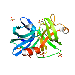
3M7U
DownloadVisualize
BU of 3m7u by Molmil
Crystal Structure of Alpha-Lytic Protease SB1+2 R64A/E182Q Mutant
Descriptor: Alpha-lytic protease, SELF-PROTEOLYSIS PRODUCT (RESIDUES 184-187), SULFATE ION
Authors:Agard, D.A, Erciyas Bailey, F.P, Waddling, C.A.
Deposit date:2010-03-17
Release date:2011-02-09
Last modified:2023-09-06
Method:X-RAY DIFFRACTION (1.05 Å)
Cite:Quantifying protein unfolding cooperativity with acid sensitive probes: Interdomain salt bridge contributions to unfolding cooperativity are combined efficiently in alpha-Lytic Protease
To be Published
3M7T
DownloadVisualize
BU of 3m7t by Molmil
Crystal Structure of Alpha-Lytic Protease SB2+3 E8A/R105S Mutant
Descriptor: Alpha-lytic protease, GLYCEROL, SULFATE ION
Authors:Agard, D.A, Erciyas Bailey, F.P, Waddling, C.A.
Deposit date:2010-03-17
Release date:2011-02-09
Last modified:2023-09-06
Method:X-RAY DIFFRACTION (1.55 Å)
Cite:Quantifying protein unfolding cooperativity with acid sensitive probes: Interdomain salt bridge contributions to unfolding cooperativity are combined efficiently in alpha-Lytic Protease
To be Published

227344

数据于2024-11-13公开中

PDB statisticsPDBj update infoContact PDBjnumon