Loading
PDBj
MenuPDBj@FacebookPDBj@TwitterPDBj@YouTubewwPDB FoundationwwPDB
RCSB PDBPDBeBMRBAdv. SearchSearch help
Search by PDB author
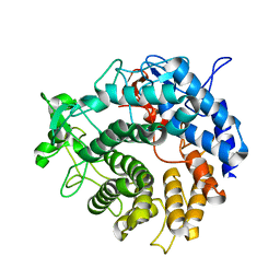
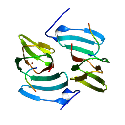
2JDA
DownloadVisualize
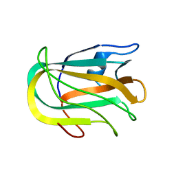
BU of 2jda by Molmil
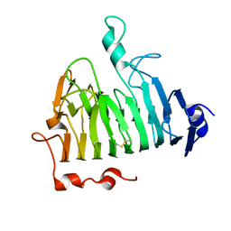
Structure of a pectin binding carbohydrate binding module determined in an monoclinic crystal form.
Descriptor: CALCIUM ION, GLYCEROL, SULFATE ION, ...
Authors:Abbott, D.W, Boraston, A.B.
Deposit date:2007-01-05
Release date:2007-01-16
Last modified:2024-05-08
Method:X-RAY DIFFRACTION (1.35 Å)
Cite:Identification and Characterization of a Novel Periplasmic Polygalacturonic Acid Binding Protein from Yersinia Enterolitica
J.Mol.Biol., 367, 2007
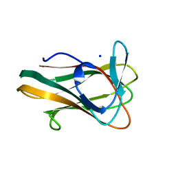
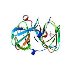
2JD9
DownloadVisualize
BU of 2jd9 by Molmil
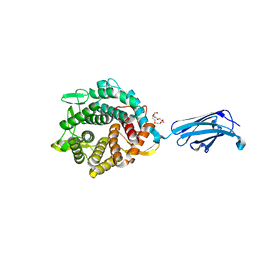
Structure of a pectin binding carbohydrate binding module determined in an orthorhombic crystal form.
Descriptor: CALCIUM ION, YECBM32
Authors:Abbott, D.W, Boraston, A.B.
Deposit date:2007-01-05
Release date:2007-01-16
Last modified:2024-05-08
Method:X-RAY DIFFRACTION (1.8 Å)
Cite:Identification and Characterization of a Novel Periplasmic Polygalacturonic Acid Binding Protein from Yersinia Enterolitica
J.Mol.Biol., 367, 2007
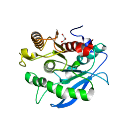
2XD3
DownloadVisualize
BU of 2xd3 by Molmil
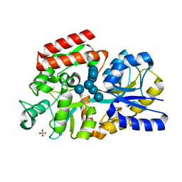
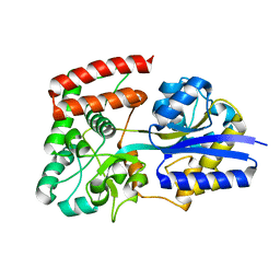
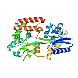
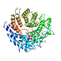
The crystal structure of MalX from Streptococcus pneumoniae in complex with maltopentaose.
Descriptor: MALTOSE/MALTODEXTRIN-BINDING PROTEIN, SULFATE ION, alpha-D-glucopyranose-(1-4)-alpha-D-glucopyranose-(1-4)-alpha-D-glucopyranose-(1-4)-alpha-D-glucopyranose-(1-4)-alpha-D-glucopyranose-(1-4)-alpha-D-glucopyranose-(1-4)-alpha-D-glucopyranose
Authors:Abbott, D.W, Higgins, M.A, Hyrnuik, S, Pluvinage, B, Lammerts van Bueren, A, Boraston, A.B.
Deposit date:2010-04-28
Release date:2010-06-09
Last modified:2023-12-20
Method:X-RAY DIFFRACTION (2 Å)
Cite:The Molecular Basis of Glycogen Breakdown and Transport in Streptococcus Pneumoniae.
Mol.Microbiol., 77, 2010
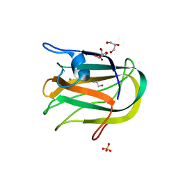
2XD2
DownloadVisualize
BU of 2xd2 by Molmil
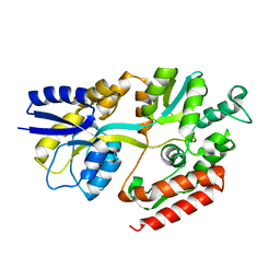
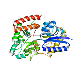
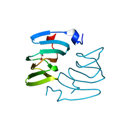
The crystal structure of MalX from Streptococcus pneumoniae
Descriptor: MALTOSE/MALTODEXTRIN-BINDING PROTEIN
Authors:Abbott, D.W, Higgins, M.A, Hyrnuik, S, Pluvinage, B, Lammerts van Bueren, A, Boraston, A.B.
Deposit date:2010-04-28
Release date:2010-06-09
Last modified:2024-05-08
Method:X-RAY DIFFRACTION (2.9 Å)
Cite:The Molecular Basis of Glycogen Breakdown and Transport in Streptococcus Pneumoniae.
Mol.Microbiol., 77, 2010
2XQX
DownloadVisualize
BU of 2xqx by Molmil
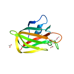
Structure of the family 32 carbohydrate-binding module from Streptococcus pneumoniae EndoD
Descriptor: ACETATE ION, CALCIUM ION, ENDO-BETA-N-ACETYLGLUCOSAMINIDASE D
Authors:Abbott, D.W, Boraston, A.B.
Deposit date:2010-09-07
Release date:2010-11-03
Last modified:2024-10-23
Method:X-RAY DIFFRACTION (2.01 Å)
Cite:Structural Analysis of a Putative Family 32 Carbohydrate-Binding Module from the Streptococcus Pneumoniae Enzyme Endod.
Acta Crystallogr.,Sect.F, 67, 2011
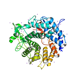
2UVE
DownloadVisualize
BU of 2uve by Molmil
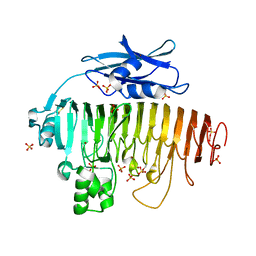
Structure of Yersinia enterocolitica Family 28 Exopolygalacturonase
Descriptor: ACETATE ION, EXOPOLYGALACTURONASE, NICKEL (II) ION, ...
Authors:Abbott, D.W, Boraston, A.B.
Deposit date:2007-03-09
Release date:2007-05-08
Last modified:2011-07-13
Method:X-RAY DIFFRACTION (2.19 Å)
Cite:The Structural Basis for Exopolygalacturonase Activity in a Family 28 Glycoside Hydrolase.
J.Mol.Biol., 368, 2007
2UVI
DownloadVisualize
BU of 2uvi by Molmil
Structure of a periplasmic oligogalacturonide binding protein from Yersinia enterocolitica in complex with 4,5-unsaturated digalacturonic acid
Descriptor: 4-deoxy-beta-L-threo-hex-4-enopyranuronic acid-(1-4)-alpha-D-galactopyranuronic acid, ABC TYPE PERIPLASMIC SUGAR-BINDING PROTEIN
Authors:Abbott, D.W, Boraston, A.B.
Deposit date:2007-03-10
Release date:2007-03-20
Last modified:2024-05-08
Method:X-RAY DIFFRACTION (2.3 Å)
Cite:Specific Recognition of Saturated and 4,5-Unsaturated Hexuronate Sugars by a Periplasmic Binding Protein Involved in Pectin Catabolism.
J.Mol.Biol., 369, 2007
2UVJ
DownloadVisualize
BU of 2uvj by Molmil
Structure of a periplasmic oligogalacturonide binding protein from Yersinia enterocolitica in complex with trigalacturonic acid
Descriptor: ABC TYPE PERIPLASMIC SUGAR-BINDING PROTEIN, alpha-D-galactopyranuronic acid-(1-4)-alpha-D-galactopyranuronic acid-(1-4)-alpha-D-galactopyranuronic acid
Authors:Abbott, D.W, Boraston, A.B.
Deposit date:2007-03-10
Release date:2007-05-22
Last modified:2024-05-08
Method:X-RAY DIFFRACTION (1.8 Å)
Cite:Specific Recognition of Saturated and 4,5-Unsaturated Hexuronate Sugars by a Periplasmic Binding Protein Involved in Pectin Catabolism.
J.Mol.Biol., 369, 2007
2UVF
DownloadVisualize
BU of 2uvf by Molmil
Structure of Yersinia enterocolitica Family 28 Exopolygalacturonase in Complex with Digalaturonic Acid
Descriptor: ACETATE ION, DI(HYDROXYETHYL)ETHER, EXOPOLYGALACTURONASE, ...
Authors:Abbott, D.W, Boraston, A.B.
Deposit date:2007-03-09
Release date:2007-05-08
Last modified:2024-10-23
Method:X-RAY DIFFRACTION (2.1 Å)
Cite:The Structural Basis for Exopolygalacturonase Activity in a Family 28 Glycoside Hydrolase.
J.Mol.Biol., 368, 2007
2UVH
DownloadVisualize
BU of 2uvh by Molmil
Structure of a periplasmic oligogalacturonide binding protein from Yersinia enterocolitica in complex with saturated digalacturonic acid
Descriptor: ABC TYPE PERIPLASMIC SUGAR-BINDING PROTEIN, alpha-D-galactopyranuronic acid-(1-4)-alpha-D-galactopyranuronic acid
Authors:Abbott, D.W, Boraston, A.B.
Deposit date:2007-03-10
Release date:2007-03-20
Last modified:2024-05-08
Method:X-RAY DIFFRACTION (2.2 Å)
Cite:Specific Recognition of Saturated and 4,5-Unsaturated Hexuronate Sugars by a Periplasmic Binding Protein Involved in Pectin Catabolism.
J.Mol.Biol., 369, 2007
2UVG
DownloadVisualize
BU of 2uvg by Molmil
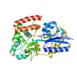
Structure of a periplasmic oligogalacturonide binding protein from Yersinia enterocolitica
Descriptor: ABC TYPE PERIPLASMIC SUGAR-BINDING PROTEIN
Authors:Abbott, D.W, Boraston, A.B.
Deposit date:2007-03-10
Release date:2007-05-22
Last modified:2024-05-08
Method:X-RAY DIFFRACTION (2.2 Å)
Cite:Specific Recognition of Saturated and 4,5-Unsaturated Hexuronate Sugars by a Periplasmic Binding Protein Involved in Pectin Catabolism.
J.Mol.Biol., 369, 2007
2W91
DownloadVisualize
BU of 2w91 by Molmil
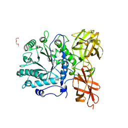
Structure of a Streptococcus pneumoniae family 85 glycoside hydrolase, Endo-D.
Descriptor: ENDO-BETA-N-ACETYLGLUCOSAMINIDASE D, PENTAETHYLENE GLYCOL
Authors:Abbott, D.W, Macauley, M.S, Vocadlo, D.J, Boraston, A.B.
Deposit date:2009-01-21
Release date:2009-01-27
Last modified:2024-05-08
Method:X-RAY DIFFRACTION (1.4 Å)
Cite:Streptococcus Pneumoniae Endohexosaminidase D, Structural and Mechanistic Insight Into Substrate-Assisted Catalysis in Family 85 Glycoside Hydrolases.
J.Biol.Chem., 284, 2009
2V8J
DownloadVisualize
BU of 2v8j by Molmil
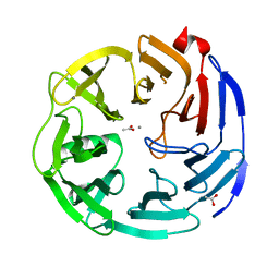
Structure of a Family 2 Pectate Lyase in Complex with a Transition Metal
Descriptor: MANGANESE (II) ION, PECTATE LYASE
Authors:Abbott, D.W, Boraston, A.B.
Deposit date:2007-08-08
Release date:2007-09-18
Last modified:2024-05-08
Method:X-RAY DIFFRACTION (2.01 Å)
Cite:A Family 2 Pectate Lyase Displays a Rare Fold and Transition Metal-Assisted -Elimination.
J.Biol.Chem., 282, 2007
2W92
DownloadVisualize
BU of 2w92 by Molmil
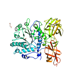
Structure of a Streptococcus pneumoniae family 85 glycoside hydrolase, Endo-D, in complex with NAG-thiazoline.
Descriptor: 3AR,5R,6S,7R,7AR-5-HYDROXYMETHYL-2-METHYL-5,6,7,7A-TETRAHYDRO-3AH-PYRANO[3,2-D]THIAZOLE-6,7-DIOL, ENDO-BETA-N-ACETYLGLUCOSAMINIDASE D, PENTAETHYLENE GLYCOL
Authors:Abbott, D.W, Macauley, M.S, Vocadlo, D.J, Boraston, A.B.
Deposit date:2009-01-21
Release date:2009-01-27
Last modified:2024-05-08
Method:X-RAY DIFFRACTION (1.65 Å)
Cite:Streptococcus Pneumoniae Endohexosaminidase D, Structural and Mechanistic Insight Into Substrate-Assisted Catalysis in Family 85 Glycoside Hydrolases.
J.Biol.Chem., 284, 2009
2V8K
DownloadVisualize
BU of 2v8k by Molmil
Structure of a Family 2 Pectate Lyase in Complex with Trigalacturonic Acid
Descriptor: PECTATE LYASE, beta-D-galactopyranuronic acid-(1-4)-alpha-D-galactopyranuronic acid-(1-4)-alpha-D-galactopyranuronic acid
Authors:Abbott, D.W, Boraston, A.B.
Deposit date:2007-08-08
Release date:2007-09-18
Last modified:2024-05-08
Method:X-RAY DIFFRACTION (2.1 Å)
Cite:A Family 2 Pectate Lyase Displays a Rare Fold and Transition Metal-Assisted Beta-Elimination.
J.Biol.Chem., 282, 2007
2V4V
DownloadVisualize
BU of 2v4v by Molmil
Crystal Structure of a Family 6 Carbohydrate-Binding Module from Clostridium cellulolyticum in complex with xylose
Descriptor: GH59 GALACTOSIDASE, SODIUM ION, beta-D-xylopyranose
Authors:Abbott, D.W, Ficko-Blean, E, Lammerts van Bueren, A, Coutinho, P.M, Henrissat, B, Gilbert, H.J, Boraston, A.B.
Deposit date:2008-09-29
Release date:2009-10-13
Last modified:2023-12-13
Method:X-RAY DIFFRACTION (1.5 Å)
Cite:Analysis of the Structural and Functional Diversity of Plant Cell Wall Specific Family 6 Carbohydrate Binding Modules.
Biochemistry, 48, 2009
2V8I
DownloadVisualize
BU of 2v8i by Molmil
Structure of a Family 2 Pectate Lyase in a Native Form
Descriptor: IODIDE ION, PECTATE LYASE
Authors:Abbott, D.W, Boraston, A.B.
Deposit date:2007-08-08
Release date:2007-09-18
Last modified:2024-05-08
Method:X-RAY DIFFRACTION (1.5 Å)
Cite:A Family 2 Pectate Lyase Displays a Rare Fold and Transition Metal-Assisted -Elimination.
J.Biol.Chem., 282, 2007
3PE7
DownloadVisualize
BU of 3pe7 by Molmil
Oligogalacturonate lyase in complex with manganese
Descriptor: ACETATE ION, CALCIUM ION, MANGANESE (II) ION, ...
Authors:Abbott, D.W, Gilbert, H.J, Boraston, A.B.
Deposit date:2010-10-25
Release date:2010-11-03
Last modified:2023-09-06
Method:X-RAY DIFFRACTION (1.65 Å)
Cite:The active site of oligogalacturonate lyase provides unique insights into cytoplasmic oligogalacturonate beta-elimination.
J.Biol.Chem., 285, 2010
3UW0
DownloadVisualize
BU of 3uw0 by Molmil
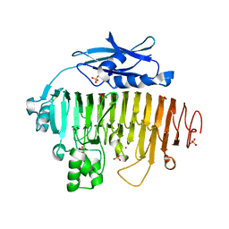
Pectin methylesterase from Yersinia enterocolitica
Descriptor: pectinesterase
Authors:Abbott, D.W, Boraston, A.B.
Deposit date:2011-11-30
Release date:2012-02-08
Last modified:2023-09-13
Method:X-RAY DIFFRACTION (3.5 Å)
Cite:Structure of a pectin methylesterase from Yersinia enterocolitica.
Acta Crystallogr.,Sect.F, 68, 2012
6U4Z
DownloadVisualize
BU of 6u4z by Molmil
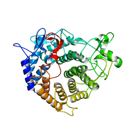
Crystal Structure of a family 76 glycoside hydrolase from a bovine Bacteroides thetaiotaomicron strain
Descriptor: 1,2-ETHANEDIOL, Alpha-1,6-mannanase, HEXAETHYLENE GLYCOL
Authors:Jones, D.R, Abbott, D.W.
Deposit date:2019-08-26
Release date:2020-04-29
Last modified:2023-10-11
Method:X-RAY DIFFRACTION (1.4 Å)
Cite:Analysis of Active Site Architecture and Reaction Product Linkage Chemistry Reveals a Conserved Cleavage Substrate for an Endo-alpha-mannanase within Diverse Yeast Mannans.
J.Mol.Biol., 432, 2020
5A29
DownloadVisualize
BU of 5a29 by Molmil
Family 2 Pectate Lyase from Vibrio vulnificus
Descriptor: 1,2-ETHANEDIOL, EXOPOLYGALACTURONATE LYASE, MANGANESE (II) ION, ...
Authors:McLean, R, Hobbs, J.K, Suits, M.D, Tuomivaara, S, Jones, D, Boraston, A.B, Abbott, D.W.
Deposit date:2015-05-15
Release date:2015-07-01
Last modified:2024-01-10
Method:X-RAY DIFFRACTION (1.9 Å)
Cite:Functional Analyses of Resurrected and Contemporary Enzymes Illuminate an Evolutionary Path for the Emergence of Exolysis in Polysaccharide Lyase Family 2.
J.Biol.Chem., 290, 2015
5FPZ
DownloadVisualize
BU of 5fpz by Molmil
The structure of KdgF from Yersinia enterocolitica with malonate bound in the active site.
Descriptor: MALONIC ACID, NICKEL (II) ION, PECTIN DEGRADATION PROTEIN
Authors:Hobbs, J.K, Lee, S.M, Robb, M, Hof, F, Barr, C, Abe, K.T, Hehemann, J.H, McLean, R, Abbott, D.W, Boraston, A.B.
Deposit date:2015-12-03
Release date:2016-05-04
Last modified:2024-01-10
Method:X-RAY DIFFRACTION (1.5 Å)
Cite:Kdgf, the Missing Link in the Microbial Metabolism of Uronate Sugars from Pectin and Alginate.
Proc.Natl.Acad.Sci.USA, 113, 2016
5FPX
DownloadVisualize
BU of 5fpx by Molmil
The structure of KdgF from Yersinia enterocolitica.
Descriptor: NICKEL (II) ION, PECTIN DEGRADATION PROTEIN, PEPTIDE
Authors:Hobbs, J.K, Lee, S.M, Robb, M, Hof, F, Barr, C, Abe, K.T, Hehemann, J.H, McLean, R, Abbott, D.W, Boraston, A.B.
Deposit date:2015-12-03
Release date:2016-05-04
Last modified:2024-01-10
Method:X-RAY DIFFRACTION (1.5 Å)
Cite:Kdgf, the Missing Link in the Microbial Metabolism of Uronate Sugars from Pectin and Alginate.
Proc.Natl.Acad.Sci.USA, 113, 2016
5FQ0
DownloadVisualize
BU of 5fq0 by Molmil
The structure of KdgF from Halomonas sp.
Descriptor: CITRATE ANION, KDGF, NICKEL (II) ION, ...
Authors:Hobbs, J.K, Lee, S.M, Robb, M, Hof, F, Barr, C, Abe, K.T, Hehemann, J.H, McLean, R, Abbott, D.W, Boraston, A.B.
Deposit date:2015-12-03
Release date:2016-05-04
Last modified:2024-01-10
Method:X-RAY DIFFRACTION (2 Å)
Cite:Kdgf, the Missing Link in the Microbial Metabolism of Uronate Sugars from Pectin and Alginate.
Proc.Natl.Acad.Sci.USA, 113, 2016
5CXU
DownloadVisualize
BU of 5cxu by Molmil
Structure of the CE1 ferulic acid esterase AmCE1/Fae1A, from the anaerobic fungi Anaeromyces mucronatus in the absence of substrate
Descriptor: GLYCEROL, ferulic acid esterase AmCE1/Fae1A
Authors:Gruninger, R.J, Abbott, D.W.
Deposit date:2015-07-29
Release date:2016-04-27
Last modified:2023-09-27
Method:X-RAY DIFFRACTION (1.6 Å)
Cite:Contributions of a unique beta-clamp to substrate recognition illuminates the molecular basis of exolysis in ferulic acid esterases.
Biochem.J., 473, 2016

226707

数据于2024-10-30公开中

PDB statisticsPDBj update infoContact PDBjnumon