Loading
PDBj
MenuPDBj@FacebookPDBj@TwitterPDBj@YouTubewwPDB FoundationwwPDB
RCSB PDBPDBeBMRBAdv. SearchSearch help
Search by PDB author
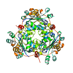
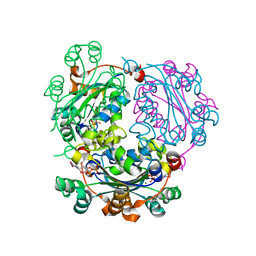
3K2C
DownloadVisualize
BU of 3k2c by Molmil
Crystal structure of peptidyl-prolyl cis-trans isomerase from Encephalitozoon cuniculi at 1.9 A resolution
Descriptor: 1,2-ETHANEDIOL, 1-METHOXY-2-[2-(2-METHOXY-ETHOXY]-ETHANE, Peptidyl-prolyl cis-trans isomerase, ...
Authors:SSGCID, Seattle Structural Genomics Center for Infectious Disease (SSGCID)
Deposit date:2009-09-29
Release date:2009-10-13
Last modified:2023-09-06
Method:X-RAY DIFFRACTION (1.95 Å)
Cite:Crystal structure of peptidyl-prolyl cis-trans isomerase from Encephalitozoon cuniculi at 1.9 A resolution
To be published
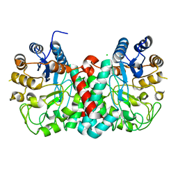
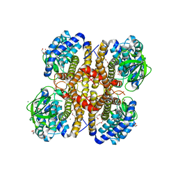
4G6C
DownloadVisualize
BU of 4g6c by Molmil
Crystal structure of beta-hexosaminidase 1 from Burkholderia cenocepacia J2315
Descriptor: Beta-hexosaminidase 1
Authors:Seattle Structural Genomics Center for Infectious Disease (SSGCID)
Deposit date:2012-07-18
Release date:2012-08-01
Last modified:2024-02-28
Method:X-RAY DIFFRACTION (1.38 Å)
Cite:Crystal structure of beta-hexosaminidase 1 from Burkholderia cenocepacia J2315
To be Published
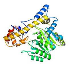
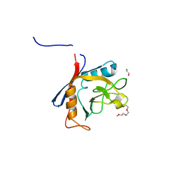
4F36
DownloadVisualize
BU of 4f36 by Molmil
Crystal structure of Nucleoside diphosphate kinase B from Trypanosoma brucei, apo form
Descriptor: Nucleoside diphosphate kinase, THIOCYANATE ION
Authors:Seattle Structural Genomics Center for Infectious Disease (SSGCID)
Deposit date:2012-05-08
Release date:2012-05-16
Last modified:2023-09-13
Method:X-RAY DIFFRACTION (2.3 Å)
Cite:Crystal structure of Nucleoside diphosphate kinase B from Trypanosoma brucei, apo form
To be Published
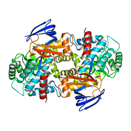
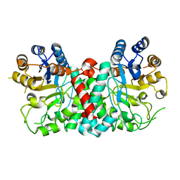
4GNV
DownloadVisualize
BU of 4gnv by Molmil
Crystal structure of beta-hexosaminidase 1 from Burkholderia cenocepacia J2315 with bound N-Acetyl-D-Glucosamine
Descriptor: 2-acetamido-2-deoxy-beta-D-glucopyranose, Beta-hexosaminidase 1, CHLORIDE ION
Authors:SSGCID, Seattle Structural Genomics Center for Infectious Disease (SSGCID)
Deposit date:2012-08-17
Release date:2012-08-29
Last modified:2024-02-28
Method:X-RAY DIFFRACTION (1.5 Å)
Cite:Crystal structure of beta-hexosaminidase 1 from Burkholderia cenocepacia J2315 with bound N-Acetyl-D-Glucosamine
TO BE PUBLISHED
4EFF
DownloadVisualize
BU of 4eff by Molmil
Crystal structure of aromatic-amino-acid aminotransferase from Burkholderia pseudomallei
Descriptor: Aromatic-amino-acid aminotransferase, GLYCEROL
Authors:Seattle Structural Genomics Center for Infectious Disease (SSGCID)
Deposit date:2012-03-29
Release date:2012-04-11
Last modified:2023-09-13
Method:X-RAY DIFFRACTION (1.85 Å)
Cite:Crystal structure of aromatic-amino-acid aminotransferase from Burkholderia pseudomallei
To be Published
4ED9
DownloadVisualize
BU of 4ed9 by Molmil
Crystal structure of a CAIB/BAIF family protein from Brucella suis
Descriptor: 1,2-ETHANEDIOL, 2-[N-CYCLOHEXYLAMINO]ETHANE SULFONIC ACID, CAIB/BAIF family protein, ...
Authors:Seattle Structural Genomics Center for Infectious Disease (SSGCID)
Deposit date:2012-03-27
Release date:2012-04-25
Last modified:2024-02-28
Method:X-RAY DIFFRACTION (1.95 Å)
Cite:Crystal structure of a CAIB/BAIF family protein from Brucella suis
To be Published
4F0L
DownloadVisualize
BU of 4f0l by Molmil
Crystal structure of Amidohydrolase from Brucella melitensis
Descriptor: Amidohydrolase, FE (III) ION, FORMIC ACID
Authors:Seattle Structural Genomics Center for Infectious Disease (SSGCID)
Deposit date:2012-05-04
Release date:2012-07-25
Last modified:2023-09-13
Method:X-RAY DIFFRACTION (2.05 Å)
Cite:Crystal structure of Amidohydrolase from Brucella melitensis
To be Published
4F4A
DownloadVisualize
BU of 4f4a by Molmil
Crystal structure of Nucleoside diphosphate kinase B from Trypanosoma brucei, UDP-bound form
Descriptor: MAGNESIUM ION, Nucleoside diphosphate kinase, URIDINE-5'-DIPHOSPHATE
Authors:Seattle Structural Genomics Center for Infectious Disease (SSGCID)
Deposit date:2012-05-10
Release date:2012-05-30
Last modified:2024-02-28
Method:X-RAY DIFFRACTION (2.1 Å)
Cite:Crystal structure of Nucleoside diphosphate kinase B from Trypanosoma brucei, UDP-bound form
To be Published
3II9
DownloadVisualize
BU of 3ii9 by Molmil
Crystal structure of glutaryl-coa dehydrogenase from Burkholderia pseudomallei at 1.73 Angstrom
Descriptor: DI(HYDROXYETHYL)ETHER, GLYCEROL, Glutaryl-CoA dehydrogenase, ...
Authors:Ismagilov, R.F, Li, L, Du, W.B, Staker, B, Accelerated Technologies Center for Gene to 3D Structure (ATCG3D), Seattle Structural Genomics Center for Infectious Disease (SSGCID)
Deposit date:2009-07-31
Release date:2009-12-15
Last modified:2023-09-06
Method:X-RAY DIFFRACTION (1.74 Å)
Cite:User-loaded SlipChip for equipment-free multiplexed nanoliter-scale experiments.
J.Am.Chem.Soc., 132, 2010
<1234


224201

数据于2024-08-28公开中

PDB statisticsPDBj update infoContact PDBjnumon