Loading
PDBj
MenuPDBj@FacebookPDBj@TwitterPDBj@YouTubewwPDB FoundationwwPDB
RCSB PDBPDBeBMRBAdv. SearchSearch help
Search by PDB author
3L11
DownloadVisualize
BU of 3l11 by Molmil
Crystal Structure of the Ring Domain of RNF168
Descriptor: E3 ubiquitin-protein ligase RNF168, MALONATE ION, ZINC ION
Authors:Neculai, D, Yermekbayeva, L, Crombet, L, Weigelt, J, Bountra, C, Edwards, A.M, Arrowsmith, C.H, Bochkarev, A, Dhe-Paganon, S, Structural Genomics Consortium (SGC)
Deposit date:2009-12-10
Release date:2010-01-19
Last modified:2023-09-06
Method:X-RAY DIFFRACTION (2.12 Å)
Cite:Molecular insights into the function of RING finger (RNF)-containing proteins hRNF8 and hRNF168 in Ubc13/Mms2-dependent ubiquitylation.
J.Biol.Chem., 287, 2012
2F9J
DownloadVisualize
BU of 2f9j by Molmil
3.0 angstrom resolution structure of a Y22M mutant of the spliceosomal protein p14 bound to a region of SF3b155
Descriptor: Pre-mRNA branch site protein p14, Splicing factor 3B subunit 1
Authors:Schellenberg, M.J, MacMillan, A.M.
Deposit date:2005-12-06
Release date:2006-01-24
Last modified:2023-08-30
Method:X-RAY DIFFRACTION (3 Å)
Cite:Crystal structure of a core spliceosomal protein interface
Proc.Natl.Acad.Sci.Usa, 103, 2006
1PZU
DownloadVisualize
BU of 1pzu by Molmil
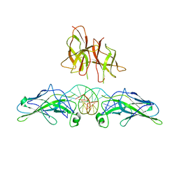
An asymmetric NFAT1-RHR homodimer on a pseudo-palindromic, Kappa-B site
Descriptor: 5'-D(*AP*AP*TP*GP*GP*AP*AP*AP*TP*TP*CP*CP*TP*C)-3', 5'-D(*TP*TP*GP*AP*GP*GP*AP*AP*TP*TP*TP*CP*CP*A)-3', Nuclear factor of activated T-cells, ...
Authors:Jin, L, Sliz, P, Chen, L, Macian, F, Rao, A, Hogan, P.G, Harrison, S.C.
Deposit date:2003-07-14
Release date:2003-09-09
Last modified:2024-02-21
Method:X-RAY DIFFRACTION (3.1 Å)
Cite:An asymmetric NFAT1 dimer on a pseudo-palindromic KB-like DNA site
Nat.Struct.Biol., 10, 2003
<1234


221716

数据于2024-06-26公开中

PDB statisticsPDBj update infoContact PDBjnumon