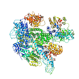
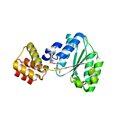
Loading
PDBj
MenuPDBj@FacebookPDBj@TwitterPDBj@YouTubewwPDB FoundationwwPDB
RCSB PDBPDBeBMRBAdv. SearchSearch help
Search by PDB author
2CHG
DownloadVisualize
BU of 2chg by Molmil
Replication Factor C domains 1 and 2
Descriptor: MAGNESIUM ION, PHOSPHOAMINOPHOSPHONIC ACID-ADENYLATE ESTER, REPLICATION FACTOR C SMALL SUBUNIT
Authors:Seybert, A, Singleton, M.R, Cook, N, Hall, D.R, Wigley, D.B.
Deposit date:2006-03-14
Release date:2006-06-06
Last modified:2024-05-08
Method:X-RAY DIFFRACTION (2.1 Å)
Cite:Communication between Subunits within an Archaeal Clamp-Loader Complex.
Embo J., 25, 2006
6S1L
DownloadVisualize
BU of 6s1l by Molmil
Structure of fission yeast Mis16
Descriptor: Histone acetyltransferase type B subunit 2
Authors:Lefevre, S, Korntner-Vetter, M, Singleton, M.R.
Deposit date:2019-06-19
Release date:2019-08-07
Last modified:2024-01-24
Method:X-RAY DIFFRACTION (1.94 Å)
Cite:Subunit interactions and arrangements in the fission yeast Mis16-Mis18-Mis19 complex.
Life Sci Alliance, 2, 2019
6S1R
DownloadVisualize
BU of 6s1r by Molmil
Structure of fission yeast Mis16 bound to histone H4
Descriptor: Histone H4, Histone acetyltransferase type B subunit 2
Authors:Lefevre, S, Korntner-Vetter, M, Singleton, M.R.
Deposit date:2019-06-19
Release date:2019-08-07
Last modified:2024-01-24
Method:X-RAY DIFFRACTION (1.8 Å)
Cite:Subunit interactions and arrangements in the fission yeast Mis16-Mis18-Mis19 complex.
Life Sci Alliance, 2, 2019
6S29
DownloadVisualize
BU of 6s29 by Molmil
Structure of fission yeast Mis16-Mis19 complex
Descriptor: BROMIDE ION, CENP-A recruiting complex protein mis19, Histone acetyltransferase type B subunit 2
Authors:Lefevre, S, Korntner-Vetter, M, Singleton, M.R.
Deposit date:2019-06-20
Release date:2019-08-07
Last modified:2024-01-24
Method:X-RAY DIFFRACTION (1.988 Å)
Cite:Subunit interactions and arrangements in the fission yeast Mis16-Mis18-Mis19 complex.
Life Sci Alliance, 2, 2019
1A2Z
DownloadVisualize
BU of 1a2z by Molmil
PYRROLIDONE CARBOXYL PEPTIDASE FROM THERMOCOCCUS LITORALIS
Descriptor: PYRROLIDONE CARBOXYL PEPTIDASE, SULFATE ION
Authors:Singleton, M.R, Isupov, M.N, Littlechild, J.A.
Deposit date:1998-01-13
Release date:1998-07-15
Last modified:2011-07-13
Method:X-RAY DIFFRACTION (1.73 Å)
Cite:X-ray structure of pyrrolidone carboxyl peptidase from the hyperthermophilic archaeon Thermococcus litoralis.
Structure Fold.Des., 7, 1999
3K70
DownloadVisualize
BU of 3k70 by Molmil
Crystal structure of the complete initiation complex of RecBCD
Descriptor: CALCIUM ION, DNA (46-MER), Exodeoxyribonuclease V alpha chain, ...
Authors:Saikrishnan, K, Wigley, D.B.
Deposit date:2009-10-11
Release date:2009-11-10
Last modified:2023-09-06
Method:X-RAY DIFFRACTION (3.59 Å)
Cite:DNA binding to RecD: role of the 1B domain in SF1B helicase activity.
Embo J., 27, 2008
1TAE
DownloadVisualize
BU of 1tae by Molmil
Structural rearrangement accompanying NAD+ synthesis within a bacterial DNA ligase crystal
Descriptor: DNA ligase, NAD-dependent, NICOTINAMIDE-ADENINE-DINUCLEOTIDE, ...
Authors:Gajiwala, K.S, Pinko, C.
Deposit date:2004-05-19
Release date:2004-11-23
Last modified:2023-08-23
Method:X-RAY DIFFRACTION (2.7 Å)
Cite:Structural rearrangement accompanying NAD+ synthesis within a bacterial DNA ligase crystal.
STRUCTURE, 12, 2004
1TA8
DownloadVisualize
BU of 1ta8 by Molmil
Structural rearrangement accompanying NAD+ synthesis within a bacterial DNA ligase crystal
Descriptor: BETA-NICOTINAMIDE RIBOSE MONOPHOSPHATE, DNA ligase, NAD-dependent, ...
Authors:Gajiwala, K.S, Pinko, C.
Deposit date:2004-05-19
Release date:2004-11-23
Last modified:2023-08-23
Method:X-RAY DIFFRACTION (1.8 Å)
Cite:Structural rearrangement accompanying NAD+ synthesis within a bacterial DNA ligase crystal.
STRUCTURE, 12, 2004
<12


224572

数据于2024-09-04公开中

PDB statisticsPDBj update infoContact PDBjnumon