Loading
PDBj
MenuPDBj@FacebookPDBj@TwitterPDBj@YouTubewwPDB FoundationwwPDB
RCSB PDBPDBeBMRBAdv. SearchSearch help
Search by PDB author
7EXE
DownloadVisualize
BU of 7exe by Molmil
Crystal structure of mouse 14-3-3zeta in complex with doubly phosphorylated ADAM22 peptide
Descriptor: 14-3-3 protein zeta/delta, Disintegrin and metalloproteinase domain-containing protein 22
Authors:Yan, L, Okatsu, K, Fukai, S.
Deposit date:2021-05-27
Release date:2021-11-24
Last modified:2023-11-29
Method:X-RAY DIFFRACTION (2.75 Å)
Cite:LGI1-ADAM22 levels regulate seizure thresholds through 14-3-3 in mice
Cell Rep, 2021
2FKE
DownloadVisualize
BU of 2fke by Molmil
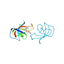
FK-506-BINDING PROTEIN: THREE-DIMENSIONAL STRUCTURE OF THE COMPLEX WITH THE ANTAGONIST L-685,818
Descriptor: 8-DEETHYL-8-[BUT-3-ENYL]-ASCOMYCIN, FK506 BINDING PROTEIN
Authors:Becker, J.W, Mckeever, B.M, Rotonda, J.
Deposit date:1993-01-27
Release date:1994-01-31
Last modified:2024-02-14
Method:X-RAY DIFFRACTION (1.72 Å)
Cite:FK-506-binding protein: three-dimensional structure of the complex with the antagonist L-685,818.
J.Biol.Chem., 268, 1993
4L35
DownloadVisualize
BU of 4l35 by Molmil
Crystal structure of cruxrhodopsin-3 at pH5 from Haloarcula vallismortis at 2.1 angstrom resolution
Descriptor: BACTERIORUBERIN, Cruxrhodopsin-3, RETINAL
Authors:Kouyama, T, Chan, S.K.
Deposit date:2013-06-05
Release date:2014-06-11
Last modified:2023-11-08
Method:X-RAY DIFFRACTION (2.1 Å)
Cite:Crystal structure of Cruxrhodopsin-3 from Haloarcula vallismortis
Plos One, 9, 2014
4JR8
DownloadVisualize
BU of 4jr8 by Molmil
Crystal structure of cruxrhodopsin-3 from Haloarcula vallismortis at 2.3 angstrom resolution
Descriptor: BACTERIORUBERIN, Cruxrhodopsin-3, RETINAL
Authors:Kouyama, T, Chan, S.K.
Deposit date:2013-03-21
Release date:2014-03-26
Last modified:2023-11-08
Method:X-RAY DIFFRACTION (2.3 Å)
Cite:Crystal structure of Cruxrhodopsin-3 from Haloarcula vallismortis
Plos One, 9, 2014
<12


224931

数据于2024-09-11公开中

PDB statisticsPDBj update infoContact PDBjnumon