Loading
PDBj
MenuPDBj@FacebookPDBj@X(formerly Twitter)PDBj@BlueSkyPDBj@YouTubewwPDB FoundationwwPDB
RCSB PDBPDBeBMRBAdv. SearchSearch help
Search by PDB author
6KP3
DownloadVisualize
BU of 6kp3 by Molmil
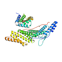
STRUCTURE OF SENDAI VIRUS Y3/ALIX-BRO1 DOMAIN COMPLEX
Descriptor: C' protein, Programmed cell death 6-interacting protein
Authors:Oda, K, Matoba, Y, Sakaguchi, T.
Deposit date:2019-08-14
Release date:2020-08-19
Last modified:2023-11-22
Method:X-RAY DIFFRACTION (2.2 Å)
Cite:Structural Insight into the Interaction of Sendai Virus C Protein with Alix To Stimulate Viral Budding.
J.Virol., 2021
3WWT
DownloadVisualize
BU of 3wwt by Molmil
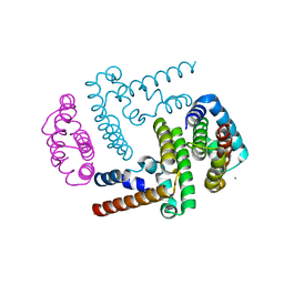
Crystal Structure of the Y3:STAT1ND complex
Descriptor: C' protein, CALCIUM ION, Signal transducer and activator of transcription 1-alpha/beta
Authors:Oda, K, Sakaguchi, T, Matoba, Y.
Deposit date:2014-06-27
Release date:2015-07-01
Last modified:2023-11-08
Method:X-RAY DIFFRACTION (2 Å)
Cite:Structural Basis of the Inhibition of STAT1 Activity by Sendai Virus C Protein.
J.Virol., 89, 2015
8I5I
DownloadVisualize
BU of 8i5i by Molmil
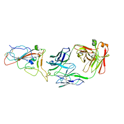
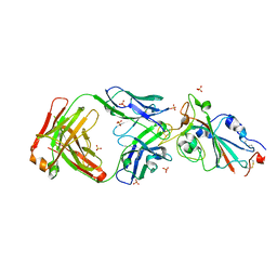
Crystal structure of SARS-CoV-2 delta variant spike receptor-binding domain (RBD) in complex with NCV2SG53 Fab
Descriptor: Fab Heavy chain, Fab Light chain, Spike protein S1
Authors:Yamamoto, A, Higashiura, A.
Deposit date:2023-01-25
Release date:2023-04-19
Last modified:2024-10-30
Method:X-RAY DIFFRACTION (3.06 Å)
Cite:Structural basis of spike RBM-specific human antibodies counteracting broad SARS-CoV-2 variants.
Commun Biol, 6, 2023
8I5H
DownloadVisualize
BU of 8i5h by Molmil
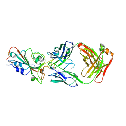
Crystal structure of SARS-CoV-2 delta variant spike receptor-binding domain (RBD) in complex with NCV2SG48 Fab
Descriptor: 2-acetamido-2-deoxy-beta-D-glucopyranose, Fab Heavy chain, Fab Light chain, ...
Authors:Yamamoto, A, Higashiura, A.
Deposit date:2023-01-25
Release date:2023-04-19
Method:X-RAY DIFFRACTION (2.38 Å)
Cite:Structural basis of spike RBM-specific human antibodies counteracting broad SARS-CoV-2 variants.
Commun Biol, 6, 2023
7WNB
DownloadVisualize
BU of 7wnb by Molmil
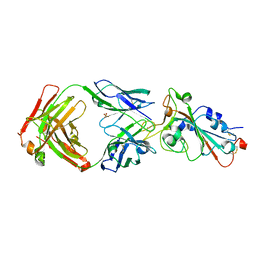
Crystal structure of SARS-CoV-2 spike receptor-binding domain (RBD) in complex with NCV2SG48 Fab
Descriptor: 2-acetamido-2-deoxy-beta-D-glucopyranose-(1-4)-2-acetamido-2-deoxy-beta-D-glucopyranose, Fab Heavy chain, Fab Light chain, ...
Authors:Yamamoto, A, Higashiura, A.
Deposit date:2022-01-18
Release date:2023-04-19
Last modified:2024-11-06
Method:X-RAY DIFFRACTION (2.18 Å)
Cite:Structural basis of spike RBM-specific human antibodies counteracting broad SARS-CoV-2 variants.
Commun Biol, 6, 2023
7WN2
DownloadVisualize
BU of 7wn2 by Molmil
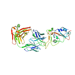
Crystal structure of SARS-CoV-2 spike receptor-binding domain (RBD) in complex with NCV2SG53 Fab
Descriptor: 2-acetamido-2-deoxy-beta-D-glucopyranose, Fab Heavy chain, Fab Light chain, ...
Authors:Yamamoto, A, Higashiura, A.
Deposit date:2022-01-17
Release date:2023-04-19
Last modified:2024-10-16
Method:X-RAY DIFFRACTION (2.35 Å)
Cite:Structural basis of spike RBM-specific human antibodies counteracting broad SARS-CoV-2 variants.
Commun Biol, 6, 2023
7YOW
DownloadVisualize
BU of 7yow by Molmil
Crystal structure of SARS-CoV-2 omicron variant spike receptor-binding domain (RBD) in complex with NCV2SG48 Fab
Descriptor: Fab Heavy chain, Fab Light chain, SULFATE ION, ...
Authors:Yamamoto, A, Higashiura, A.
Deposit date:2022-08-02
Release date:2023-04-19
Last modified:2024-11-13
Method:X-RAY DIFFRACTION (3.3 Å)
Cite:Structural basis of spike RBM-specific human antibodies counteracting broad SARS-CoV-2 variants.
Commun Biol, 6, 2023
7VDY
DownloadVisualize
BU of 7vdy by Molmil
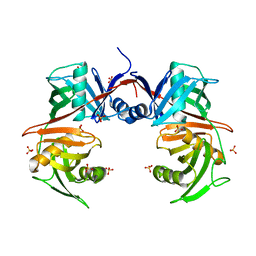
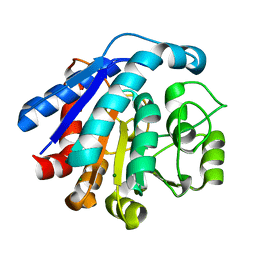
Crystal structure of O-ureidoserine racemase
Descriptor: O-ureido-serine racemase, SULFATE ION
Authors:Oda, K, Matoba, Y.
Deposit date:2021-09-07
Release date:2021-12-15
Last modified:2023-11-29
Method:X-RAY DIFFRACTION (2.12 Å)
Cite:Crystal structure of O-ureidoserine racemase found in the d-cycloserine biosynthetic pathway.
Proteins, 90, 2022
7EUN
DownloadVisualize
BU of 7eun by Molmil
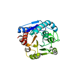
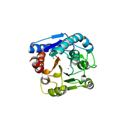
Crystal structure of N(omega)-hydroxy-L-arginine hydrolase in complex with ABH
Descriptor: 2(S)-AMINO-6-BORONOHEXANOIC ACID, MAGNESIUM ION, MANGANESE (II) ION, ...
Authors:Oda, K, Matoba, Y.
Deposit date:2021-05-18
Release date:2022-05-18
Last modified:2023-11-29
Method:X-RAY DIFFRACTION (1.28 Å)
Cite:Catalytic mechanism of DcsB: Arginase framework used for hydrolyzing its inhibitor.
Protein Sci., 31, 2022
7EUQ
DownloadVisualize
BU of 7euq by Molmil
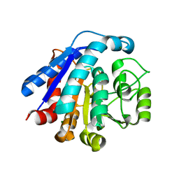
Crystal structure of C86H-Y124N-G126H-H196S mutant of N(omega)-hydroxy-L-arginine hydrolase
Descriptor: MAGNESIUM ION, MANGANESE (II) ION, N(omega)-hydroxy-L-arginine amidinohydrolase
Authors:Oda, K, Matoba, Y.
Deposit date:2021-05-18
Release date:2022-05-18
Last modified:2023-11-29
Method:X-RAY DIFFRACTION (1.8 Å)
Cite:Catalytic mechanism of DcsB: Arginase framework used for hydrolyzing its inhibitor.
Protein Sci., 31, 2022
7EUL
DownloadVisualize
BU of 7eul by Molmil
Crystal structure of C86H-H196S mutant of N(omega)-hydroxy-L-arginine hydrolase
Descriptor: MAGNESIUM ION, MANGANESE (II) ION, N(omega)-hydroxy-L-arginine amidinohydrolase
Authors:Oda, K, Matoba, Y.
Deposit date:2021-05-18
Release date:2022-05-18
Last modified:2023-11-29
Method:X-RAY DIFFRACTION (1.45 Å)
Cite:Catalytic mechanism of DcsB: Arginase framework used for hydrolyzing its inhibitor.
Protein Sci., 31, 2022
7EUK
DownloadVisualize
BU of 7euk by Molmil
Crystal structure of N(omega)-hydroxy-L-arginine hydrolase in complex with L-Orn
Descriptor: L-ornithine, MAGNESIUM ION, MANGANESE (II) ION, ...
Authors:Oda, K, Matoba, Y.
Deposit date:2021-05-18
Release date:2022-05-18
Last modified:2023-11-29
Method:X-RAY DIFFRACTION (1.4 Å)
Cite:Catalytic mechanism of DcsB: Arginase framework used for hydrolyzing its inhibitor.
Protein Sci., 31, 2022

227344

건을2024-11-13부터공개중

PDB statisticsPDBj update infoContact PDBjnumon