Loading
PDBj
MenuPDBj@FacebookPDBj@TwitterPDBj@YouTubewwPDB FoundationwwPDB
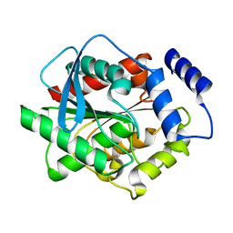
RCSB PDBPDBeBMRBAdv. SearchSearch help
Search by PDB author
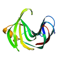
8WXP
DownloadVisualize
BU of 8wxp by Molmil
Crystal structure of substrate-binding protein from Rhodothermus marinus (Dose IV)
Descriptor: ABC-type uncharacterized transport system periplasmic component-like protein
Authors:Nam, K.H.
Deposit date:2023-10-30
Release date:2023-11-22
Last modified:2024-05-22
Method:X-RAY DIFFRACTION (1.75 Å)
Cite:Data of radiation damage on selenomethionine-substituted single-domain substrate-binding protein.
Data Brief, 53, 2024
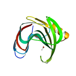
8WXM
DownloadVisualize
BU of 8wxm by Molmil
Crystal structure of substrate-binding protein from Rhodothermus marinus (Dose I)
Descriptor: ABC-type uncharacterized transport system periplasmic component-like protein
Authors:Nam, K.H.
Deposit date:2023-10-30
Release date:2023-11-22
Last modified:2024-05-22
Method:X-RAY DIFFRACTION (1.5 Å)
Cite:Data of radiation damage on selenomethionine-substituted single-domain substrate-binding protein.
Data Brief, 53, 2024
8WDH
DownloadVisualize
BU of 8wdh by Molmil
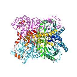
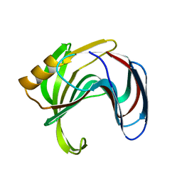
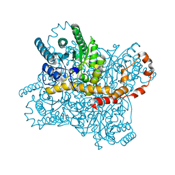
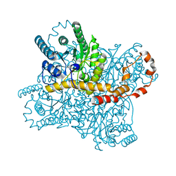
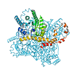
Crystal structure of glucose isomerase by fixed-target pink-beam serial synchrotron crystallography
Descriptor: MAGNESIUM ION, Xylose isomerase
Authors:Nam, K.H.
Deposit date:2023-09-15
Release date:2023-11-29
Last modified:2024-04-17
Method:X-RAY DIFFRACTION (1.7 Å)
Cite:Fixed-Target Pink-Beam Serial Synchrotron Crystallography at Pohang Light Source II.
Crystals, 13, 2023
8WGP
DownloadVisualize
BU of 8wgp by Molmil
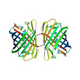
Crystal structure of DsRed-Monomer
Descriptor: Red fluorescent protein
Authors:Nam, K.H.
Deposit date:2023-09-22
Release date:2024-02-21
Method:X-RAY DIFFRACTION (2.9 Å)
Cite:Structural Flexibility of the Monomeric Red Fluorescent Protein DsRed.
Crystals, 14, 2024
8HVF
DownloadVisualize
BU of 8hvf by Molmil
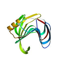
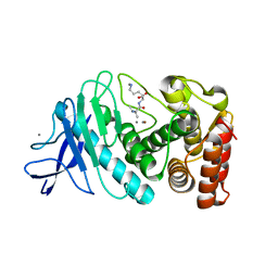
Crystal structure of Thaumatin (100 ms)
Descriptor: 1,2-ETHANEDIOL, L(+)-TARTARIC ACID, Thaumatin I
Authors:Nam, K.H.
Deposit date:2022-12-26
Release date:2023-11-08
Method:X-RAY DIFFRACTION (1.13 Å)
Cite:Crystal structure of Thaumatin (100 ms)
To Be Published
8HVE
DownloadVisualize
BU of 8hve by Molmil
Crystal structure of Thaumatin (1 s)
Descriptor: 1,2-ETHANEDIOL, L(+)-TARTARIC ACID, Thaumatin I
Authors:Nam, K.H.
Deposit date:2022-12-26
Release date:2023-11-08
Method:X-RAY DIFFRACTION (1.13 Å)
Cite:Crystal structure of Thaumatin (1 s)
To Be Published
6LZO
DownloadVisualize
BU of 6lzo by Molmil
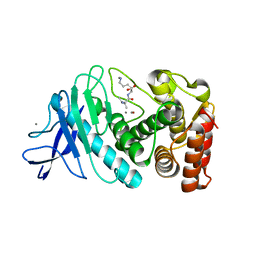
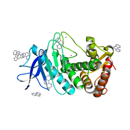
Thermolysin with 1,10-phenanthroline
Descriptor: 1,10-PHENANTHROLINE, CALCIUM ION, Thermolysin
Authors:Nam, K.H.
Deposit date:2020-02-19
Release date:2021-01-27
Last modified:2023-11-29
Method:X-RAY DIFFRACTION (1.8 Å)
Cite:Structural analysis of metal chelation of the metalloproteinase thermolysin by 1,10-phenanthroline.
J.Inorg.Biochem., 215, 2021
3L1J
DownloadVisualize
BU of 3l1j by Molmil
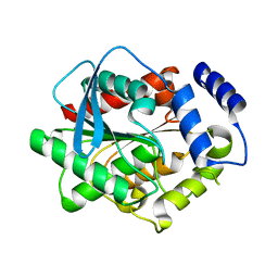
Crystal structure of EstE5, was soaked by ZnSO4
Descriptor: Esterase/lipase
Authors:Nam, K.H, Hwang, K.Y.
Deposit date:2009-12-11
Release date:2010-01-19
Last modified:2023-11-01
Method:X-RAY DIFFRACTION (2 Å)
Cite:Structural insights into the noninvasive inhibition of HSL-homolog EstE5 by organic solvents and metal ions
To be Published
3L1H
DownloadVisualize
BU of 3l1h by Molmil
Crystal structure of EstE5, was soaked by FeCl3
Descriptor: Esterase/lipase
Authors:Nam, K.H, Hwang, K.Y.
Deposit date:2009-12-11
Release date:2010-01-19
Last modified:2023-11-01
Method:X-RAY DIFFRACTION (2.4 Å)
Cite:Structural insights into the noninvasive inhibition of HSL-homolog EstE5 by organic solvents and metal ions
To be Published
3L1I
DownloadVisualize
BU of 3l1i by Molmil
Crystal structure of EstE5, was soaked by CuSO4
Descriptor: Esterase/lipase
Authors:Nam, K.H, Hwang, K.Y.
Deposit date:2009-12-11
Release date:2010-01-19
Last modified:2023-11-01
Method:X-RAY DIFFRACTION (2.2 Å)
Cite:Structural insights into the noninvasive inhibition of HSL-homolog EstE5 by organic solvents and metal ions
To be Published
7BVL
DownloadVisualize
BU of 7bvl by Molmil
Crystal structure of glucose isomerase delivered in wheat starch
Descriptor: MAGNESIUM ION, Xylose isomerase
Authors:Nam, K.H.
Deposit date:2020-04-11
Release date:2020-11-18
Last modified:2023-11-29
Method:X-RAY DIFFRACTION (2 Å)
Cite:Polysaccharide-Based Injection Matrix for Serial Crystallography.
Int J Mol Sci, 21, 2020
7BVO
DownloadVisualize
BU of 7bvo by Molmil
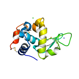
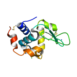
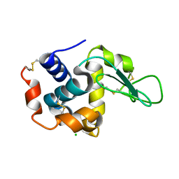
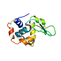
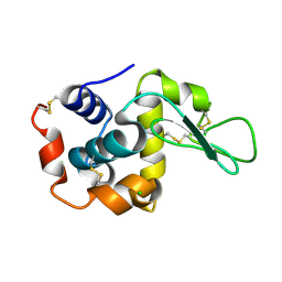
Crystal structure of lysozyme delivered in alginate
Descriptor: CHLORIDE ION, Lysozyme C
Authors:Nam, K.H.
Deposit date:2020-04-11
Release date:2020-11-18
Last modified:2023-11-29
Method:X-RAY DIFFRACTION (1.9 Å)
Cite:Polysaccharide-Based Injection Matrix for Serial Crystallography.
Int J Mol Sci, 21, 2020
7BVM
DownloadVisualize
BU of 7bvm by Molmil
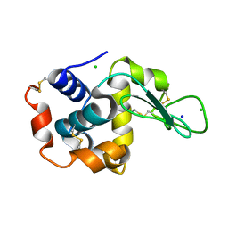
Crystal structure of lysozyme delivered in wheat starch
Descriptor: CHLORIDE ION, Lysozyme C
Authors:Nam, K.H.
Deposit date:2020-04-11
Release date:2020-11-18
Last modified:2023-11-29
Method:X-RAY DIFFRACTION (2 Å)
Cite:Polysaccharide-Based Injection Matrix for Serial Crystallography.
Int J Mol Sci, 21, 2020
7BVN
DownloadVisualize
BU of 7bvn by Molmil
Crystal structure of glucose isomerase delivered in alginate
Descriptor: MAGNESIUM ION, Xylose isomerase
Authors:Nam, K.H.
Deposit date:2020-04-11
Release date:2020-11-18
Last modified:2023-11-29
Method:X-RAY DIFFRACTION (2 Å)
Cite:Polysaccharide-Based Injection Matrix for Serial Crystallography.
Int J Mol Sci, 21, 2020
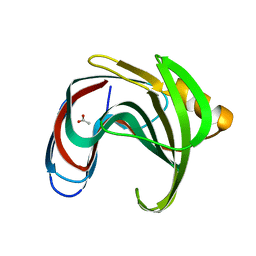
8WXN
DownloadVisualize
BU of 8wxn by Molmil
Crystal structure of substrate-binding protein from Rhodothermus marinus (Dose II)
Descriptor: ABC-type uncharacterized transport system periplasmic component-like protein
Authors:Nam, K.H.
Deposit date:2023-10-30
Release date:2023-11-22
Last modified:2024-05-22
Method:X-RAY DIFFRACTION (1.58 Å)
Cite:Data of radiation damage on selenomethionine-substituted single-domain substrate-binding protein.
Data Brief, 53, 2024
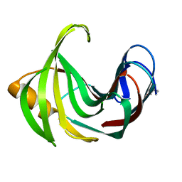
8WXO
DownloadVisualize
BU of 8wxo by Molmil
Crystal structure of substrate-binding protein from Rhodothermus marinus (Dose III)
Descriptor: ABC-type uncharacterized transport system periplasmic component-like protein
Authors:Nam, K.H.
Deposit date:2023-10-30
Release date:2023-11-22
Last modified:2024-05-22
Method:X-RAY DIFFRACTION (1.68 Å)
Cite:Data of radiation damage on selenomethionine-substituted single-domain substrate-binding protein.
Data Brief, 53, 2024
6LZN
DownloadVisualize
BU of 6lzn by Molmil
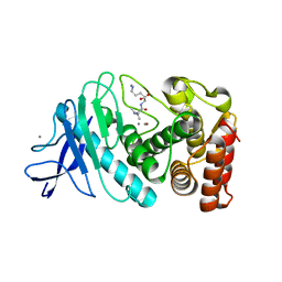
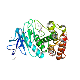
Thermolysin
Descriptor: CALCIUM ION, GLYCEROL, ISOLEUCINE, ...
Authors:Nam, K.H.
Deposit date:2020-02-19
Release date:2021-01-27
Last modified:2023-11-29
Method:X-RAY DIFFRACTION (1.5 Å)
Cite:Structural analysis of metal chelation of the metalloproteinase thermolysin by 1,10-phenanthroline.
J.Inorg.Biochem., 215, 2021
6LL2
DownloadVisualize
BU of 6ll2 by Molmil
Crystal structure of glucose isomerase by fixed-target serial femtosecond crystallography
Descriptor: MAGNESIUM ION, Xylose isomerase
Authors:Nam, K.H.
Deposit date:2019-12-21
Release date:2020-06-10
Last modified:2023-11-22
Method:X-RAY DIFFRACTION (1.75 Å)
Cite:Crystal structure of glucose isomerase by fixed-target serial femtosecond crystallography
To Be Published
6LL3
DownloadVisualize
BU of 6ll3 by Molmil
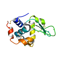
Crystal structure of lysozyme by fixed-target serial femtosecond crystallography
Descriptor: CHLORIDE ION, Lysozyme C
Authors:Nam, K.H.
Deposit date:2019-12-21
Release date:2020-06-10
Last modified:2023-11-22
Method:X-RAY DIFFRACTION (1.85 Å)
Cite:Crystal structure of lysozyme by fixed-target serial femtosecond crystallography
To Be Published
8WDI
DownloadVisualize
BU of 8wdi by Molmil
Crystal structure of lysozyme by fixed-target pink-beam serial synchrotron crystallography
Descriptor: CHLORIDE ION, Lysozyme C
Authors:Nam, K.H.
Deposit date:2023-09-15
Release date:2023-10-04
Last modified:2024-04-17
Method:X-RAY DIFFRACTION (2.2 Å)
Cite:Fixed-Target Pink-Beam Serial Synchrotron Crystallography at Pohang Light Source II.
Crystals, 13, 2023
8WFV
DownloadVisualize
BU of 8wfv by Molmil
Crystal structure of beta-glucosidase from Thermoanaerobacterium saccharolyticum (Data 3)
Descriptor: 2-AMINO-2-HYDROXYMETHYL-PROPANE-1,3-DIOL, Beta-glucosidase, SODIUM ION
Authors:Nam, K.H.
Deposit date:2023-09-20
Release date:2023-10-04
Last modified:2024-04-17
Method:X-RAY DIFFRACTION (1.5 Å)
Cite:The Conformational Change of the L3 Loop Affects the Structural Changes in the Substrate Binding Pocket Entrance of beta-Glucosidase.
Molecules, 28, 2023
8WGL
DownloadVisualize
BU of 8wgl by Molmil
Crystal structure of Rhodothermus marinus substrate-binding protein (Hg soaking)
Descriptor: ABC-type uncharacterized transport system periplasmic component-like protein, MERCURY (II) ION
Authors:Nam, K.H.
Deposit date:2023-09-22
Release date:2023-10-04
Last modified:2024-04-17
Method:X-RAY DIFFRACTION (2.3 Å)
Cite:Structural and bioinformatics analysis of single-domain substrate-binding protein from Rhodothermus marinus.
Biochem Biophys Rep, 37, 2024
8WFT
DownloadVisualize
BU of 8wft by Molmil
Crystal structure of beta-glucosidase from Thermoanaerobacterium saccharolyticum (Data 1)
Descriptor: 1,2-ETHANEDIOL, 2-AMINO-2-HYDROXYMETHYL-PROPANE-1,3-DIOL, Beta-glucosidase, ...
Authors:Nam, K.H.
Deposit date:2023-09-20
Release date:2023-10-04
Last modified:2024-04-17
Method:X-RAY DIFFRACTION (1.9 Å)
Cite:The Conformational Change of the L3 Loop Affects the Structural Changes in the Substrate Binding Pocket Entrance of beta-Glucosidase.
Molecules, 28, 2023
8WGK
DownloadVisualize
BU of 8wgk by Molmil
Crystal structure of Rhodothermus marinus substrate-binding protein (Br soaking)
Descriptor: ABC-type uncharacterized transport system periplasmic component-like protein
Authors:Nam, K.H.
Deposit date:2023-09-22
Release date:2023-10-04
Last modified:2024-04-17
Method:X-RAY DIFFRACTION (1.75 Å)
Cite:Structural and bioinformatics analysis of single-domain substrate-binding protein from Rhodothermus marinus.
Biochem Biophys Rep, 37, 2024
8WFW
DownloadVisualize
BU of 8wfw by Molmil
Crystal structure of beta-glucosidase from Thermoanaerobacterium saccharolyticum (Data 4)
Descriptor: 2-AMINO-2-HYDROXYMETHYL-PROPANE-1,3-DIOL, Beta-glucosidase, SODIUM ION
Authors:Nam, K.H.
Deposit date:2023-09-20
Release date:2023-10-04
Last modified:2024-04-17
Method:X-RAY DIFFRACTION (1.61 Å)
Cite:The Conformational Change of the L3 Loop Affects the Structural Changes in the Substrate Binding Pocket Entrance of beta-Glucosidase.
Molecules, 28, 2023

221051

건을2024-06-12부터공개중

PDB statisticsPDBj update infoContact PDBjnumon