Loading
PDBj
MenuPDBj@FacebookPDBj@TwitterPDBj@YouTubewwPDB FoundationwwPDB
RCSB PDBPDBeBMRBAdv. SearchSearch help
Search by PDB author
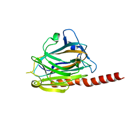
3POS
DownloadVisualize
BU of 3pos by Molmil
Crystal structure of the globular domain of human calreticulin
Descriptor: CALCIUM ION, Calreticulin
Authors:Chouquet, A, Paidassi, H, Ling, W.-L, Frachet, P, Houen, G, Arlaud, G.J, Gaboriaud, C.
Deposit date:2010-11-23
Release date:2011-03-09
Last modified:2024-10-16
Method:X-RAY DIFFRACTION (1.65 Å)
Cite:X-ray structure of the human calreticulin globular domain reveals a Peptide-binding area and suggests a multi-molecular mechanism
Plos One, 6, 2011
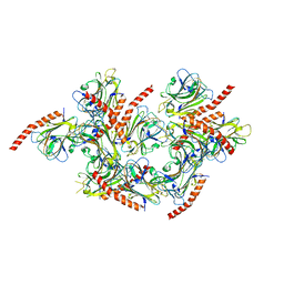
3POW
DownloadVisualize
BU of 3pow by Molmil
Crystal structure of the globular domain of human calreticulin
Descriptor: CALCIUM ION, calreticulin
Authors:Gaboriaud, C.
Deposit date:2010-11-23
Release date:2011-03-09
Last modified:2024-10-30
Method:X-RAY DIFFRACTION (1.55 Å)
Cite:X-ray structure of the human calreticulin globular domain reveals a Peptide-binding area and suggests a multi-molecular mechanism
Plos One, 6, 2011
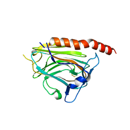
5LK5
DownloadVisualize
BU of 5lk5 by Molmil
Crystal structure of the globular domain of human calreticulin mutant D71K
Descriptor: CALCIUM ION, CHLORIDE ION, Calreticulin,Calreticulin
Authors:Gaboriaud, C, Cioci, G.
Deposit date:2016-07-21
Release date:2016-08-31
Last modified:2019-10-16
Method:X-RAY DIFFRACTION (2.3 Å)
Cite:Structures of parasite calreticulins provide insights into their flexibility and dual carbohydrate/peptide-binding properties.
IUCrJ, 3, 2016

226707

건을2024-10-30부터공개중

PDB statisticsPDBj update infoContact PDBjnumon