Loading
PDBj
MenuPDBj@FacebookPDBj@TwitterPDBj@YouTubewwPDB FoundationwwPDB
RCSB PDBPDBeBMRBAdv. SearchSearch help
Search by PDB author
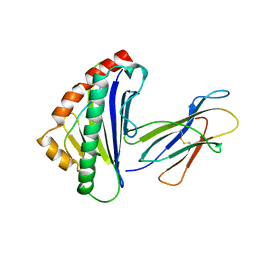
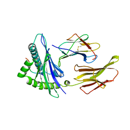
1HSB
DownloadVisualize
BU of 1hsb by Molmil
DIFFERENT LENGTH PEPTIDES BIND TO HLA-AW68 SIMILARLY AT THEIR ENDS BUT BULGE OUT IN THE MIDDLE
Descriptor: ALANINE, ARGININE, BOUND PEPTIDE FRAGMENT, ...
Authors:Guo, H.-C, Strominger, J.L, Wiley, D.C.
Deposit date:1993-03-30
Release date:1993-10-31
Last modified:2024-10-30
Method:X-RAY DIFFRACTION (1.9 Å)
Cite:Different length peptides bind to HLA-Aw68 similarly at their ends but bulge out in the middle.
Nature, 360, 1992
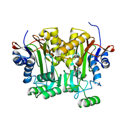
5J5E
DownloadVisualize
BU of 5j5e by Molmil
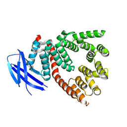
crystal structure of antigen-ERAP1 domain complex
Descriptor: Endoplasmic reticulum aminopeptidase 1
Authors:Sui, L, Gandhi, A, Guo, H.-C.
Deposit date:2016-04-02
Release date:2017-04-12
Last modified:2023-09-27
Method:X-RAY DIFFRACTION (2.8 Å)
Cite:Crystal structure of a polypeptide's C-terminus in complex with the regulatory domain of ER aminopeptidase 1.
Mol.Immunol., 80, 2016
1YFI
DownloadVisualize
BU of 1yfi by Molmil
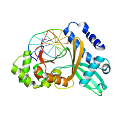
Crystal Structure of restriction endonuclease MspI in complex with its cognate DNA in P212121 space group
Descriptor: 5'-D(*CP*CP*CP*CP*CP*GP*GP*GP*GP*G)-3', Type II restriction enzyme MspI
Authors:Xu, Q.S, Kucera, R.B, Roberts, R.J, Guo, H.-C.
Deposit date:2005-01-02
Release date:2006-01-10
Last modified:2023-08-23
Method:X-RAY DIFFRACTION (2.7 Å)
Cite:Two crystal forms of the restriction enzyme MspI-DNA complex show the same novel structure.
Protein Sci., 14, 2005
3RJO
DownloadVisualize
BU of 3rjo by Molmil
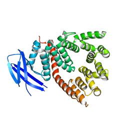
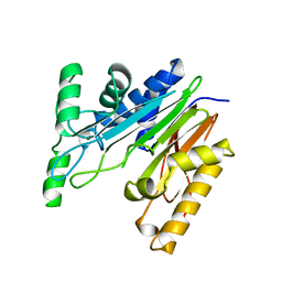
Crystal Structure of ERAP1 Peptide Binding Domain
Descriptor: 1,2-ETHANEDIOL, Endoplasmic reticulum aminopeptidase 1
Authors:Guo, H.-C, Lakshminarasimhan, D, Gandhi, A.
Deposit date:2011-04-15
Release date:2011-12-21
Last modified:2012-03-07
Method:X-RAY DIFFRACTION (2.3 Å)
Cite:Structural insights into the molecular ruler mechanism of the endoplasmic reticulum aminopeptidase ERAP1.
Sci Rep, 1, 2011
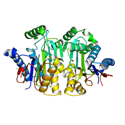
2GAC
DownloadVisualize
BU of 2gac by Molmil
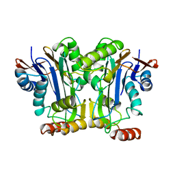
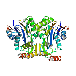
T152C MUTANT GLYCOSYLASPARAGINASE FROM FLAVOBACTERIUM MENINGOSEPTICUM
Descriptor: GLYCOSYLASPARAGINASE
Authors:Guo, H.-C, Xu, Q.
Deposit date:1998-05-29
Release date:1999-06-08
Last modified:2024-02-14
Method:X-RAY DIFFRACTION (2.1 Å)
Cite:Crystal structures of Flavobacterium glycosylasparaginase. An N-terminal nucleophile hydrolase activated by intramolecular proteolysis.
J.Biol.Chem., 273, 1998
2GAW
DownloadVisualize
BU of 2gaw by Molmil
WILD TYPE GLYCOSYLASPARAGINASE FROM FLAVOBACTERIUM MENINGOSEPTICUM
Descriptor: GLYCOSYLASPARAGINASE
Authors:Guo, H.-C, Xu, Q.
Deposit date:1998-05-29
Release date:1999-06-08
Last modified:2024-02-14
Method:X-RAY DIFFRACTION (2.2 Å)
Cite:Crystal structures of Flavobacterium glycosylasparaginase. An N-terminal nucleophile hydrolase activated by intramolecular proteolysis.
J.Biol.Chem., 273, 1998
9GAA
DownloadVisualize
BU of 9gaa by Molmil
PRECURSOR OF THE T152A MUTANT GLYCOSYLASPARAGINASE FROM FLAVOBACTERIUM MENINGOSEPTICUM
Descriptor: PROTEIN (GLYCOSYLASPARAGINASE)
Authors:Guo, H.-C, Xu, Q.
Deposit date:1999-06-15
Release date:2000-06-16
Last modified:2023-09-20
Method:X-RAY DIFFRACTION (2.1 Å)
Cite:Structural insights into the mechanism of intramolecular proteolysis.
Cell(Cambridge,Mass.), 98, 1999
9GAF
DownloadVisualize
BU of 9gaf by Molmil
PRECURSOR OF THE W11F MUTANT GLYCOSYLASPARAGINASE FROM FLAVOBACTERIUM MENINGOSEPTICUM
Descriptor: GLYCINE, PROTEIN (GLYCOSYLASPARAGINASE)
Authors:Guo, H.-C, Xu, Q.
Deposit date:1999-06-15
Release date:2000-06-16
Last modified:2023-09-20
Method:X-RAY DIFFRACTION (1.9 Å)
Cite:Structural insights into the mechanism of intramolecular proteolysis.
Cell(Cambridge,Mass.), 98, 1999
9GAC
DownloadVisualize
BU of 9gac by Molmil
PRECURSOR OF THE T152C MUTANT GLYCOSYLASPARAGINASE FROM FLAVOBACTERIUM MENINGOSEPTICUM
Descriptor: GLYCINE, PROTEIN (GLYCOSYLASPARAGINASE)
Authors:Guo, H.-C, Xu, Q.
Deposit date:1999-06-15
Release date:2000-06-16
Last modified:2023-09-20
Method:X-RAY DIFFRACTION (1.9 Å)
Cite:Structural insights into the mechanism of intramolecular proteolysis.
Cell(Cambridge,Mass.), 98, 1999
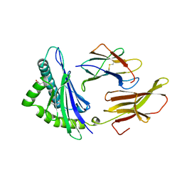
1TMC
DownloadVisualize
BU of 1tmc by Molmil
THE THREE-DIMENSIONAL STRUCTURE OF A CLASS I MAJOR HISTOCOMPATIBILITY COMPLEX MOLECULE MISSING THE ALPHA3 DOMAIN OF THE HEAVY CHAIN
Descriptor: BETA 2-MICROGLOBULIN, CLASS I HISTOCOMPATIBILITY ANTIGEN (HLA-AW68), DECAMERIC PEPTIDE (EVAPPEYHRK)
Authors:Collins, E.J, Garboczi, D.N, Karpusas, M.N, Wiley, D.C.
Deposit date:1994-12-19
Release date:1995-03-31
Last modified:2024-10-30
Method:X-RAY DIFFRACTION (2.3 Å)
Cite:The three-dimensional structure of a class I major histocompatibility complex molecule missing the alpha 3 domain of the heavy chain.
Proc.Natl.Acad.Sci.USA, 92, 1995
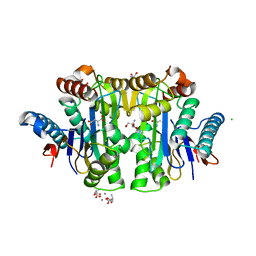
2ZAK
DownloadVisualize
BU of 2zak by Molmil
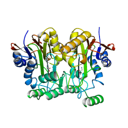
Orthorhombic crystal structure of precursor E. coli isoaspartyl peptidase/L-asparaginase (EcAIII) with active-site T179A mutation
Descriptor: 2-AMINO-2-HYDROXYMETHYL-PROPANE-1,3-DIOL, CHLORIDE ION, L-asparaginase precursor, ...
Authors:Michalska, K, Hernandez-Santoyo, A, Jaskolski, M.
Deposit date:2007-10-07
Release date:2008-03-25
Last modified:2023-11-01
Method:X-RAY DIFFRACTION (2.01 Å)
Cite:Crystal packing of plant-type L-asparaginase from Escherichia coli
Acta Crystallogr.,Sect.D, 64, 2008
2ZAL
DownloadVisualize
BU of 2zal by Molmil
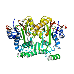
Crystal structure of E. coli isoaspartyl aminopeptidase/L-asparaginase in complex with L-aspartate
Descriptor: 2-AMINO-2-HYDROXYMETHYL-PROPANE-1,3-DIOL, ASPARTIC ACID, CALCIUM ION, ...
Authors:Michalska, K, Brzezinski, K, Jaskolski, M.
Deposit date:2007-10-07
Release date:2007-10-30
Last modified:2023-11-01
Method:X-RAY DIFFRACTION (1.9 Å)
Cite:Crystal structure of isoaspartyl aminopeptidase in complex with L-aspartate
J.Biol.Chem., 280, 2005
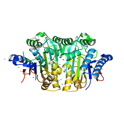
3C17
DownloadVisualize
BU of 3c17 by Molmil
Hexagonal Crystal Structure of Precursor E. coli Isoaspartyl Peptidase/l-Asparaginase (ECAIII) with Active-site T179A mutation
Descriptor: CHLORIDE ION, L-asparaginase precursor, SODIUM ION
Authors:Michalska, K, Hernandez-Santoyo, A, Jaskolski, M.
Deposit date:2008-01-22
Release date:2008-04-01
Last modified:2023-11-01
Method:X-RAY DIFFRACTION (1.95 Å)
Cite:The Mechanism of Autocatalytic Activation of Plant-type L-Asparaginases
J.Biol.Chem., 283, 2008
2CLR
DownloadVisualize
BU of 2clr by Molmil
THREE DIMENSIONAL STRUCTURE OF A PEPTIDE EXTENDING OUT ONE END OF A CLASS I MHC BINDING SITE
Descriptor: BETA 2-MICROGLOBULIN, CLASS I HISTOCOMPATIBILITY ANTIGEN (HLA-A 0201) (ALPHA CHAIN), DECAMERIC PEPTIDE FROM CALRETICULIN
Authors:Collins, E.J, Garboczi, D.N, Wiley, D.C.
Deposit date:1994-08-01
Release date:1995-03-31
Last modified:2024-10-16
Method:X-RAY DIFFRACTION (2 Å)
Cite:Three-dimensional structure of a peptide extending from one end of a class I MHC binding site.
Nature, 371, 1994
3LJQ
DownloadVisualize
BU of 3ljq by Molmil
Crystal Structure of the Glycosylasparaginase T152C apo-precursor
Descriptor: GLYCINE, N(4)-(Beta-N-acetylglucosaminyl)-L-asparaginase, SODIUM ION
Authors:Wang, Y, Guo, H.-C.
Deposit date:2010-01-26
Release date:2010-11-24
Last modified:2023-09-06
Method:X-RAY DIFFRACTION (1.9 Å)
Cite:Crystallographic snapshot of glycosylasparaginase precursor poised for autoprocessing.
J.Mol.Biol., 403, 2010

227111

건을2024-11-06부터공개중

PDB statisticsPDBj update infoContact PDBjnumon