Loading
PDBj
MenuPDBj@FacebookPDBj@X(formerly Twitter)PDBj@BlueSkyPDBj@YouTubewwPDB FoundationwwPDB
RCSB PDBPDBeBMRBAdv. SearchSearch help
Search by PDB author
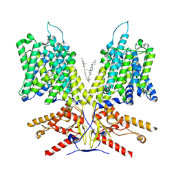
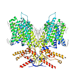
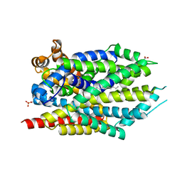
3USG
DownloadVisualize
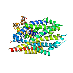
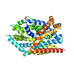
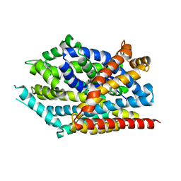
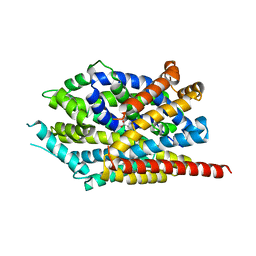
BU of 3usg by Molmil
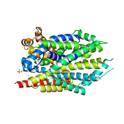
Crystal structure of LeuT bound to L-leucine in space group C2 from lipid bicelles
Descriptor: ACETATE ION, DI(HYDROXYETHYL)ETHER, LEUCINE, ...
Authors:Wang, H, Elferich, J, Gouaux, E.
Deposit date:2011-11-23
Release date:2012-01-11
Last modified:2023-09-13
Method:X-RAY DIFFRACTION (2.502 Å)
Cite:Structures of LeuT in bicelles define conformation and substrate binding in a membrane-like context.
Nat.Struct.Mol.Biol., 19, 2012
6C10
DownloadVisualize
BU of 6c10 by Molmil
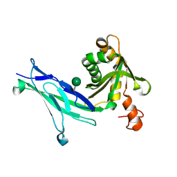
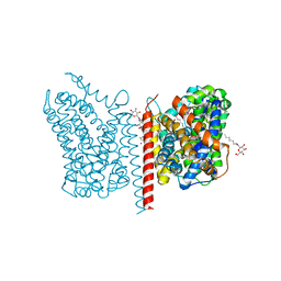
Crystal structure of mouse PCDH15 EC11-EL
Descriptor: Protocadherin-15, alpha-D-mannopyranose
Authors:Gouaux, E, Elferich, J, Ge, J.
Deposit date:2018-01-03
Release date:2018-08-15
Last modified:2024-10-16
Method:X-RAY DIFFRACTION (1.399 Å)
Cite:Structure of mouse protocadherin 15 of the stereocilia tip link in complex with LHFPL5.
Elife, 7, 2018
6C13
DownloadVisualize
BU of 6c13 by Molmil
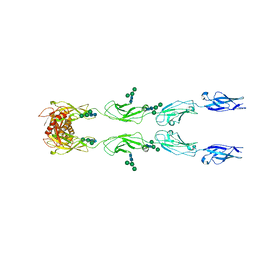
CryoEM structure of mouse PCDH15-4EC-LHFPL5 complex
Descriptor: Protocadherin-15, alpha-D-mannopyranose-(1-3)-[alpha-D-mannopyranose-(1-6)]beta-D-mannopyranose-(1-4)-2-acetamido-2-deoxy-beta-D-glucopyranose-(1-4)-2-acetamido-2-deoxy-beta-D-glucopyranose
Authors:Gouaux, E, Ge, J, Elferich, J.
Deposit date:2018-01-03
Release date:2018-08-15
Last modified:2024-10-16
Method:ELECTRON MICROSCOPY (11.33 Å)
Cite:Structure of mouse protocadherin 15 of the stereocilia tip link in complex with LHFPL5.
Elife, 7, 2018
6C14
DownloadVisualize
BU of 6c14 by Molmil
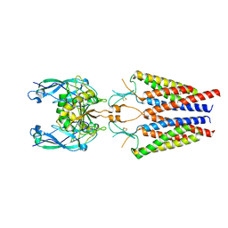
CryoEM structure of mouse PCDH15-1EC-LHFPL5 complex
Descriptor: LHFPL tetraspan subfamily member 5 protein, Protocadherin-15
Authors:Gouaux, E, Elferich, J, Ge, J.
Deposit date:2018-01-03
Release date:2018-08-15
Last modified:2019-12-18
Method:ELECTRON MICROSCOPY (4.5 Å)
Cite:Structure of mouse protocadherin 15 of the stereocilia tip link in complex with LHFPL5.
Elife, 7, 2018
3USJ
DownloadVisualize
BU of 3usj by Molmil
Crystal structure of LeuT bound to L-leucine in space group P21 from lipid bicelles
Descriptor: LEUCINE, SODIUM ION, Transporter
Authors:Wang, H, Elferich, J, Gouaux, E.
Deposit date:2011-11-23
Release date:2012-01-11
Last modified:2024-02-28
Method:X-RAY DIFFRACTION (3.5 Å)
Cite:Structures of LeuT in bicelles define conformation and substrate binding in a membrane-like context.
Nat.Struct.Mol.Biol., 19, 2012
3USL
DownloadVisualize
BU of 3usl by Molmil
Crystal Structure of LeuT bound to L-selenomethionine in space group C2 from lipid bicelles
Descriptor: ACETATE ION, IODIDE ION, PHOSPHOCHOLINE, ...
Authors:Wang, H, Elferich, J, Gouaux, E.
Deposit date:2011-11-23
Release date:2012-01-11
Last modified:2023-12-06
Method:X-RAY DIFFRACTION (2.71 Å)
Cite:Structures of LeuT in bicelles define conformation and substrate binding in a membrane-like context.
Nat.Struct.Mol.Biol., 19, 2012
3USK
DownloadVisualize
BU of 3usk by Molmil
Crystal structure of LeuT bound to L-leucine in space group P21 from lipid bicelles
Descriptor: LEUCINE, SODIUM ION, Transporter
Authors:Wang, H, Elferich, J, Gouaux, E.
Deposit date:2011-11-23
Release date:2012-01-11
Last modified:2023-09-13
Method:X-RAY DIFFRACTION (4.5 Å)
Cite:Structures of LeuT in bicelles define conformation and substrate binding in a membrane-like context.
Nat.Struct.Mol.Biol., 19, 2012
3USO
DownloadVisualize
BU of 3uso by Molmil
Crystal structure of LeuT bound to L-selenomethionine in space group P21212 from lipid bicelles
Descriptor: SELENOMETHIONINE, SODIUM ION, Transporter
Authors:Wang, H, Elferich, J, Gouaux, E.
Deposit date:2011-11-23
Release date:2012-01-11
Last modified:2023-12-06
Method:X-RAY DIFFRACTION (4.5 Å)
Cite:Structures of LeuT in bicelles define conformation and substrate binding in a membrane-like context.
Nat.Struct.Mol.Biol., 19, 2012
3USI
DownloadVisualize
BU of 3usi by Molmil
Crystal structure of LeuT bound to L-leucine in space group P2 from lipid bicelles
Descriptor: LEUCINE, SODIUM ION, Transporter
Authors:Wang, H, Elferich, J, Gouaux, E.
Deposit date:2011-11-23
Release date:2012-01-11
Last modified:2023-09-13
Method:X-RAY DIFFRACTION (3.106 Å)
Cite:Structures of LeuT in bicelles define conformation and substrate binding in a membrane-like context.
Nat.Struct.Mol.Biol., 19, 2012
3USM
DownloadVisualize
BU of 3usm by Molmil
Crystal Structure of LeuT bound to L-selenomethionine in space group C2 from lipid bicelles (collected at 1.2 A)
Descriptor: IODIDE ION, PHOSPHOCHOLINE, SELENOMETHIONINE, ...
Authors:Wang, H, Elferich, J, Gouaux, E.
Deposit date:2011-11-23
Release date:2012-01-11
Last modified:2023-12-06
Method:X-RAY DIFFRACTION (3.008 Å)
Cite:Structures of LeuT in bicelles define conformation and substrate binding in a membrane-like context.
Nat.Struct.Mol.Biol., 19, 2012
3USP
DownloadVisualize
BU of 3usp by Molmil
Crystal structure of LeuT in heptyl-beta-D-Selenoglucoside
Descriptor: CHLORIDE ION, LEUCINE, SODIUM ION, ...
Authors:Wang, H, Elferich, J, Gouaux, E.
Deposit date:2011-11-23
Release date:2012-01-11
Last modified:2023-09-13
Method:X-RAY DIFFRACTION (2.1 Å)
Cite:Structures of LeuT in bicelles define conformation and substrate binding in a membrane-like context.
Nat.Struct.Mol.Biol., 19, 2012
7LGU
DownloadVisualize
BU of 7lgu by Molmil
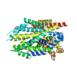
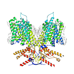
Structure of human prestin in the presence of NaCl
Descriptor: CHLORIDE ION, CHOLESTEROL, DECANE, ...
Authors:Ge, J, Gouaux, E.
Deposit date:2021-01-21
Release date:2021-08-25
Last modified:2024-05-29
Method:ELECTRON MICROSCOPY (2.3 Å)
Cite:Molecular mechanism of prestin electromotive signal amplification.
Cell, 184, 2021
7LH3
DownloadVisualize
BU of 7lh3 by Molmil
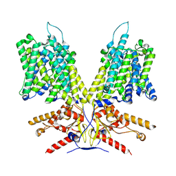
Structure of human prestin in the presence of sodium sulfate
Descriptor: Prestin
Authors:Ge, J, Gouaux, E.
Deposit date:2021-01-21
Release date:2021-08-25
Last modified:2024-05-29
Method:ELECTRON MICROSCOPY (4.3 Å)
Cite:Molecular mechanism of prestin electromotive signal amplification.
Cell, 184, 2021
7LH2
DownloadVisualize
BU of 7lh2 by Molmil
Structure of human prestin in the presence of sodium salicylate and sodium sulfate
Descriptor: 2-HYDROXYBENZOIC ACID, CHOLESTEROL, Prestin
Authors:Ge, J, Gouaux, E.
Deposit date:2021-01-21
Release date:2021-08-25
Last modified:2024-05-29
Method:ELECTRON MICROSCOPY (3.43 Å)
Cite:Molecular mechanism of prestin electromotive signal amplification.
Cell, 184, 2021
7LGW
DownloadVisualize
BU of 7lgw by Molmil
Structure of human Prestin in nanodisc in the presence of NaCl
Descriptor: CHLORIDE ION, CHOLESTEROL, DECANE, ...
Authors:Ge, J, Gouaux, E.
Deposit date:2021-01-21
Release date:2021-08-25
Last modified:2024-05-29
Method:ELECTRON MICROSCOPY (2.7 Å)
Cite:Molecular mechanism of prestin electromotive signal amplification.
Cell, 184, 2021

227344

건을2024-11-13부터공개중

PDB statisticsPDBj update infoContact PDBjnumon