Loading
PDBj
MenuPDBj@FacebookPDBj@TwitterPDBj@YouTubewwPDB FoundationwwPDB
RCSB PDBPDBeBMRBAdv. SearchSearch help
Search by PDB author
8EB9
DownloadVisualize
BU of 8eb9 by Molmil
Crystal structure of SIX8 from Fusarium oxysporum f. sp. lycopersici
Descriptor: SULFATE ION, Secreted in xylem Six8
Authors:Yu, D.S, Ericsson, D.J, Williams, S.J.
Deposit date:2022-08-30
Release date:2023-06-21
Last modified:2024-04-03
Method:X-RAY DIFFRACTION (1.28 Å)
Cite:The structural repertoire of Fusarium oxysporum f. sp. lycopersici effectors revealed by experimental and computational studies.
Elife, 12, 2024
8EBB
DownloadVisualize
BU of 8ebb by Molmil
Crystal structure of SIX6 from Fusarium oxysporum f. sp. lycopersici
Descriptor: D(-)-TARTARIC ACID, Secreted in xylem Six6
Authors:Yu, D.S, Ericsson, D.J, Williams, S.J.
Deposit date:2022-08-30
Release date:2023-06-21
Last modified:2024-04-03
Method:X-RAY DIFFRACTION (1.88 Å)
Cite:The structural repertoire of Fusarium oxysporum f. sp. lycopersici effectors revealed by experimental and computational studies.
Elife, 12, 2024
8P0E
DownloadVisualize
BU of 8p0e by Molmil
Rubella virus p150 macro domain in complex with ADP-ribose
Descriptor: ADENOSINE-5-DIPHOSPHORIBOSE, Non-structural polyprotein p200
Authors:Stoll, G.A, Modis, Y.
Deposit date:2023-05-10
Release date:2024-01-17
Last modified:2024-03-06
Method:X-RAY DIFFRACTION (1.59 Å)
Cite:Crystal structure and biochemical activity of the macrodomain from rubella virus p150.
J.Virol., 98, 2024
6EYC
DownloadVisualize
BU of 6eyc by Molmil
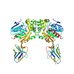
Re-refinement of the MCM2-7 double hexamer using ISOLDE
Descriptor: ADENOSINE-5'-DIPHOSPHATE, DNA replication licensing factor MCM2, DNA replication licensing factor MCM3, ...
Authors:Croll, T.I.
Deposit date:2017-11-11
Release date:2018-06-20
Last modified:2024-05-08
Method:ELECTRON MICROSCOPY (3.8 Å)
Cite:ISOLDE: a physically realistic environment for model building into low-resolution electron-density maps.
Acta Crystallogr D Struct Biol, 74, 2018
8QZO
DownloadVisualize
BU of 8qzo by Molmil
Crystal structure of heterodimeric complex of CdpB1 and CdpB2 from A. fulgidus
Descriptor: PRC-barrel domain-containing protein
Authors:Ciziene, D, Bellini, D, Lowe, J.
Deposit date:2023-10-27
Release date:2024-04-03
Method:X-RAY DIFFRACTION (2.29 Å)
Cite:Proteins containing photosynthetic reaction centre domains modulate FtsZ-based archaeal cell division.
Nat Microbiol, 9, 2024
7BBM
DownloadVisualize
BU of 7bbm by Molmil
Mutant nitrobindin M75L/H76L/Q96C/M148L (NB4H) from Arabidopsis thaliana with cofactor MnPPIX
Descriptor: 1,2-ETHANEDIOL, MANGANESE PROTOPORPHYRIN IX, UPF0678 fatty acid-binding protein-like protein At1g79260
Authors:Minges, A, Sauer, D.F, Wittwer, M, Markel, U, Spiertz, M, Schiffels, J, Davari, M.D, Okuda, J, Schwaneberg, U, Groth, G.
Deposit date:2020-12-18
Release date:2021-05-26
Last modified:2024-01-31
Method:X-RAY DIFFRACTION (1.14 Å)
Cite:Chemogenetic engineering of nitrobindin toward an artificial epoxygenase
Catalysis Science And Technology, 2021
7DZ5
DownloadVisualize
BU of 7dz5 by Molmil
Crystal structures of D-allulose 3-epimerase with D-sorbose from Sinorhizobium fredii
Descriptor: D-sorbose, D-tagatose 3-epimerase, MAGNESIUM ION, ...
Authors:Zhu, Z.L, Miyakawa, T, Tanokura, M, Lu, F.P, Qin, H.-M.
Deposit date:2021-01-23
Release date:2022-08-03
Last modified:2023-11-29
Method:X-RAY DIFFRACTION (1.7 Å)
Cite:Substantial Improvement of an Epimerase for the Synthesis of D-Allulose by Biosensor-Based High-Throughput Microdroplet Screening
Angew.Chem.Int.Ed.Engl., 2023
7QKB
DownloadVisualize
BU of 7qkb by Molmil
Crystal structure of human Cathepsin L in complex with covalently bound GC376
Descriptor: CHLORIDE ION, Cathepsin L, DI(HYDROXYETHYL)ETHER, ...
Authors:Reinke, P.Y.A, Falke, S, Lieske, J, Ewert, W, Loboda, J, Rahmani Mashhour, A, Hauser, M, Karnicar, K, Usenik, A, Lindic, N, Lach, M, Boehler, H, Beck, T, Cox, R, Chapman, H.N, Hinrichs, W, Turk, D, Guenther, S, Meents, A.
Deposit date:2021-12-17
Release date:2022-12-28
Last modified:2024-01-31
Method:X-RAY DIFFRACTION (1.8 Å)
Cite:Calpeptin is a potent cathepsin inhibitor and drug candidate for SARS-CoV-2 infections.
Commun Biol, 6, 2023
7QKA
DownloadVisualize
BU of 7qka by Molmil
Crystal structure of SARS-CoV-2 Main Protease in complex with covalently bound GC376
Descriptor: 3C-like proteinase nsp5, DIMETHYL SULFOXIDE, N~2~-[(benzyloxy)carbonyl]-N-{(2S)-1-hydroxy-3-[(3S)-2-oxopyrrolidin-3-yl]propan-2-yl}-L-leucinamide
Authors:Reinke, P.Y.A, Falke, S, Lieske, J, Ewert, W, Loboda, J, Rahmani Mashhour, A, Hauser, M, Karnicar, K, Usenik, A, Lindic, N, Lach, M, Boehler, H, Beck, T, Cox, R, Chapman, H.N, Hinrichs, W, Turk, D, Guenther, S, Meents, A.
Deposit date:2021-12-17
Release date:2022-12-28
Last modified:2024-01-31
Method:X-RAY DIFFRACTION (1.8 Å)
Cite:Calpeptin is a potent cathepsin inhibitor and drug candidate for SARS-CoV-2 infections.
Commun Biol, 6, 2023
8EE2
DownloadVisualize
BU of 8ee2 by Molmil
Crystal Structure of Nanobody VHH219 Bound to Its Antigen PA14 Cif
Descriptor: CFTR inhibitory factor, Nanobody VHH219
Authors:Simard, A.R, Madden, D.R.
Deposit date:2022-09-06
Release date:2023-09-13
Method:X-RAY DIFFRACTION (2.4 Å)
Cite:Crystal Structure of Nanobody VHH219 Bound to Its Antigen PA14 Cif
To Be Published
8E2N
DownloadVisualize
BU of 8e2n by Molmil
Crystal Structure of Nanobody VHH113 Specific for PA14 Cif
Descriptor: Nanobody VHH113
Authors:Simard, A.R, Madden, D.R.
Deposit date:2022-08-15
Release date:2023-10-04
Method:X-RAY DIFFRACTION (1.1 Å)
Cite:Crystal Structure of Nanobody VHH113 Specific for PA14 Cif
To Be Published
8F6U
DownloadVisualize
BU of 8f6u by Molmil
Crystal Structure of Nanobody VHH113 Bound to Its Antigen PA14 Cif
Descriptor: CFTR inhibitory factor, Nanobody VHH113
Authors:Simard, A.R, Taher, N.M, Mishra, A.K, Madden, D.R.
Deposit date:2022-11-17
Release date:2024-03-13
Method:X-RAY DIFFRACTION (1.7 Å)
Cite:Crystal Structure of Nanobody VHH113 Bound to Its Antigen PA14 Cif
To Be Published
8F6V
DownloadVisualize
BU of 8f6v by Molmil
Crystal Structure of Nanobody VHH108 Bound to Its Antigen PA14 Cif
Descriptor: CFTR inhibitory factor, Nanobody VHH108
Authors:Simard, A.R, Madden, D.R.
Deposit date:2022-11-17
Release date:2024-03-13
Method:X-RAY DIFFRACTION (2.3 Å)
Cite:Crystal Structure of Nanobody VHH113 Bound to Its Antigen PA14 Cif
To Be Published
8TT5
DownloadVisualize
BU of 8tt5 by Molmil
Pseudomonas fluorescens isocyanide hydratase pH=8.3
Descriptor: 1,2-ETHANEDIOL, Isonitrile hydratase InhA
Authors:Wilson, M.A, Smith, N, Dasgupta, M, Dolamore, C.
Deposit date:2023-08-12
Release date:2023-09-20
Method:X-RAY DIFFRACTION (1.02 Å)
Cite:Changes in an Enzyme Ensemble During Catalysis Observed by High Resolution XFEL Crystallography.
Biorxiv, 2023
8TSU
DownloadVisualize
BU of 8tsu by Molmil
Pseudomonas fluorescens G150T-1 isocyanide hydratase at 274 K
Descriptor: CHLORIDE ION, Isonitrile hydratase InhA
Authors:Wilson, M.A, Smith, N, Dasgupta, M, Dolamore, C.
Deposit date:2023-08-11
Release date:2023-09-20
Method:X-RAY DIFFRACTION (1.15 Å)
Cite:Changes in an Enzyme Ensemble During Catalysis Observed by High Resolution XFEL Crystallography.
Biorxiv, 2023
8TSX
DownloadVisualize
BU of 8tsx by Molmil
Pseudomonas fluorescens G150T isocyanide hydratase at 100 K
Descriptor: CHLORIDE ION, Isonitrile hydratase InhA
Authors:Wilson, M.A, Smith, N, Dasgupta, M, Dolamore, C.
Deposit date:2023-08-12
Release date:2023-09-20
Method:X-RAY DIFFRACTION (1 Å)
Cite:Changes in an Enzyme Ensemble During Catalysis Observed by High Resolution XFEL Crystallography.
Biorxiv, 2023
8TT1
DownloadVisualize
BU of 8tt1 by Molmil
Pseudomonas fluorescens isocyanide hydratase pH=5.0
Descriptor: Isonitrile hydratase InhA
Authors:Wilson, M.A, Smith, N, Dasgupta, M, Dolamore, C.
Deposit date:2023-08-12
Release date:2023-09-20
Method:X-RAY DIFFRACTION (1.45 Å)
Cite:Changes in an Enzyme Ensemble During Catalysis Observed by High Resolution XFEL Crystallography.
Biorxiv, 2023
8TSZ
DownloadVisualize
BU of 8tsz by Molmil
Pseudomonas fluorescens G150T-3 isocyanide hydratase at 274 K
Descriptor: CHLORIDE ION, Isonitrile hydratase InhA
Authors:Wilson, M.A, Smith, N, Dasgupta, M, Dolamore, C.
Deposit date:2023-08-12
Release date:2023-09-20
Method:X-RAY DIFFRACTION (1.1 Å)
Cite:Changes in an Enzyme Ensemble During Catalysis Observed by High Resolution XFEL Crystallography.
Biorxiv, 2023
8TT4
DownloadVisualize
BU of 8tt4 by Molmil
Pseudomonas fluorescens isocyanide hydratase pH=6.0
Descriptor: 1,2-ETHANEDIOL, Isonitrile hydratase InhA
Authors:Wilson, M.A, Smith, N, Dasgupta, M, Dolamore, C.
Deposit date:2023-08-12
Release date:2023-09-20
Method:X-RAY DIFFRACTION (1.2 Å)
Cite:Changes in an Enzyme Ensemble During Catalysis Observed by High Resolution XFEL Crystallography.
Biorxiv, 2023
8TSY
DownloadVisualize
BU of 8tsy by Molmil
Pseudomonas fluorescens G150T-2 isocyanide hydratase at 274 K
Descriptor: CHLORIDE ION, Isonitrile hydratase InhA
Authors:Wilson, M.A, Smith, N, Dasgupta, M, Dolamore, C.
Deposit date:2023-08-12
Release date:2023-09-20
Method:X-RAY DIFFRACTION (1.2 Å)
Cite:Changes in an Enzyme Ensemble During Catalysis Observed by High Resolution XFEL Crystallography.
Biorxiv, 2023
8TT0
DownloadVisualize
BU of 8tt0 by Molmil
Pseudomonas fluorescens isocyanide hydratase pH=4.2
Descriptor: Isonitrile hydratase InhA
Authors:Wilson, M.A, Smith, N, Dasgupta, M, Dolamore, C.
Deposit date:2023-08-12
Release date:2023-09-20
Method:X-RAY DIFFRACTION (1.5 Å)
Cite:Changes in an Enzyme Ensemble During Catalysis Observed by High Resolution XFEL Crystallography.
Biorxiv, 2023
8TT2
DownloadVisualize
BU of 8tt2 by Molmil
Pseudomonas fluorescens isocyanide hydratase pH=5.4
Descriptor: 1,2-ETHANEDIOL, Isonitrile hydratase InhA
Authors:Wilson, M.A, Smith, N, Dasgupta, M, Dolamore, C.
Deposit date:2023-08-12
Release date:2023-09-20
Method:X-RAY DIFFRACTION (1.33 Å)
Cite:Changes in an Enzyme Ensemble During Catalysis Observed by High Resolution XFEL Crystallography.
Biorxiv, 2023
6WYH
DownloadVisualize
BU of 6wyh by Molmil
Crystal structure of Human H-chain Ferritin variant 157C Delta C-star Modified with a RAFT Agent Soaked in an Acrylate Solution
Descriptor: CALCIUM ION, FE (III) ION, Ferritin heavy chain, ...
Authors:Bailey, J.B, Zhang, L.
Deposit date:2020-05-12
Release date:2020-11-11
Last modified:2023-10-18
Method:X-RAY DIFFRACTION (2.22 Å)
Cite:Anisotropic Dynamics and Mechanics of Macromolecular Crystals Containing Lattice-Patterned Polymer Networks.
J.Am.Chem.Soc., 142, 2020
6WYF
DownloadVisualize
BU of 6wyf by Molmil
Crystal structure of Human H-chain Ferritin variant 157C Delta C-star Modified with a RAFT Agent Soaked in an Acrylate Solution
Descriptor: CALCIUM ION, FE (III) ION, Ferritin heavy chain, ...
Authors:Bailey, J.B, Zhang, L.
Deposit date:2020-05-12
Release date:2020-11-11
Last modified:2023-10-18
Method:X-RAY DIFFRACTION (1.25 Å)
Cite:Anisotropic Dynamics and Mechanics of Macromolecular Crystals Containing Lattice-Patterned Polymer Networks.
J.Am.Chem.Soc., 142, 2020
6WPM
DownloadVisualize
BU of 6wpm by Molmil
Crystal structure of a putative oligosaccharide periplasmic-binding protein from Synechococcus sp. MITs9220 in complex with zinc
Descriptor: 1,2-ETHANEDIOL, GLYCEROL, SULFATE ION, ...
Authors:Ford, B.A, Michie, K.A, Paulsen, I.T, Mabbutt, B.C, Shah, B.S.
Deposit date:2020-04-27
Release date:2021-05-12
Last modified:2022-04-27
Method:X-RAY DIFFRACTION (2.88 Å)
Cite:Novel functional insights into a modified sugar-binding protein from Synechococcus MITS9220.
Sci Rep, 12, 2022

221716

건을2024-06-26부터공개중

PDB statisticsPDBj update infoContact PDBjnumon