Loading
PDBj
MenuPDBj@FacebookPDBj@TwitterPDBj@YouTubewwPDB FoundationwwPDB
RCSB PDBPDBeBMRBAdv. SearchSearch help
Search by PDB author
5SD4
DownloadVisualize
BU of 5sd4 by Molmil
Crystal Structure of Dihydrofolate Reductase from Mycobacterium tuberculosis bound to NADP and SDDC Inhibitor SDDC-1218
Descriptor: 1,2-ETHANEDIOL, 3-(2-{3-[(2,4-diamino-6-ethylpyrimidin-5-yl)oxy]propoxy}pyridin-3-yl)prop-2-yn-1-ol, CHLORIDE ION, ...
Authors:Seattle Structural Genomics Center for Infectious Disease (SSGCID)
Deposit date:2021-12-20
Release date:2022-02-09
Last modified:2024-05-22
Method:X-RAY DIFFRACTION (1.25 Å)
Cite:Crystal Structure of Dihydrofolate Reductase from Mycobacterium tuberculosis bound to NADP and SDDC Inhibitor SDDC-1218
to be published
5SD6
DownloadVisualize
BU of 5sd6 by Molmil
Crystal Structure of Dihydrofolate Reductase from Homo sapiens bound to NADP and SDDC Inhibitor SDDC-892
Descriptor: 1,2-ETHANEDIOL, 6-ethyl-5-(4-{2-[2-(1H-tetrazol-5-yl)ethyl]phenoxy}butoxy)pyrimidine-2,4-diamine, CHLORIDE ION, ...
Authors:Seattle Structural Genomics Center for Infectious Disease (SSGCID)
Deposit date:2021-12-20
Release date:2022-02-09
Last modified:2024-05-22
Method:X-RAY DIFFRACTION (2.15 Å)
Cite:Crystal Structure of Dihydrofolate Reductase from Homo sapiens bound to NADP and SDDC Inhibitor SDDC-892
to be published
5SCN
DownloadVisualize
BU of 5scn by Molmil
Crystal Structure of Dihydrofolate Reductase from Mycobacterium tuberculosis bound to NADP and SDDC Inhibitor SDDC-25
Descriptor: 3-(2-{3-[(2,4-diamino-6-ethylpyrimidin-5-yl)oxy]propoxy}phenyl)-N-(methanesulfonyl)propanamide, Dihydrofolate reductase, NADP NICOTINAMIDE-ADENINE-DINUCLEOTIDE PHOSPHATE
Authors:Seattle Structural Genomics Center for Infectious Disease (SSGCID)
Deposit date:2021-12-20
Release date:2022-02-09
Last modified:2024-05-22
Method:X-RAY DIFFRACTION (1.45 Å)
Cite:Crystal Structure of Dihydrofolate Reductase from Mycobacterium tuberculosis bound to NADP and SDDC Inhibitor SDDC-25
to be published
5SCP
DownloadVisualize
BU of 5scp by Molmil
Crystal Structure of Dihydrofolate Reductase from Mycobacterium tuberculosis bound to NADP and SDDC Inhibitor SDDC-23
Descriptor: 1,2-ETHANEDIOL, 3-(2-{3-[(2,4-diamino-6-ethylpyrimidin-5-yl)oxy]propoxy}phenyl)propanamide, CHLORIDE ION, ...
Authors:Seattle Structural Genomics Center for Infectious Disease (SSGCID)
Deposit date:2021-12-20
Release date:2022-02-09
Last modified:2024-05-22
Method:X-RAY DIFFRACTION (1.8 Å)
Cite:Crystal Structure of Dihydrofolate Reductase from Mycobacterium tuberculosis bound to NADP and SDDC Inhibitor SDDC-23
to be published
3V9P
DownloadVisualize
BU of 3v9p by Molmil
Crystal structure of Thymidylate kinase from Burkholderia thailandensis
Descriptor: (4R)-2-METHYLPENTANE-2,4-DIOL, ACETATE ION, DI(HYDROXYETHYL)ETHER, ...
Authors:Seattle Structural Genomics Center for Infectious Disease (SSGCID)
Deposit date:2011-12-27
Release date:2012-01-25
Last modified:2023-09-13
Method:X-RAY DIFFRACTION (1.9 Å)
Cite:Combining functional and structural genomics to sample the essential Burkholderia structome.
Plos One, 8, 2013
3UPT
DownloadVisualize
BU of 3upt by Molmil
Crystal structure of a transketolase from Burkholderia pseudomallei bound to TPP, calcium and ribose-5-phosphate
Descriptor: 5-O-phosphono-beta-D-ribofuranose, BROMIDE ION, CALCIUM ION, ...
Authors:Seattle Structural Genomics Center for Infectious Disease (SSGCID)
Deposit date:2011-11-18
Release date:2011-12-14
Last modified:2023-09-13
Method:X-RAY DIFFRACTION (2.4 Å)
Cite:Combining functional and structural genomics to sample the essential Burkholderia structome.
Plos One, 8, 2013
3U0I
DownloadVisualize
BU of 3u0i by Molmil
Crystal Structure of a probable FAD-binding, putative uncharacterized protein from Brucella melitensis
Descriptor: 1,2-ETHANEDIOL, 2-[N-CYCLOHEXYLAMINO]ETHANE SULFONIC ACID, probable FAD-binding, ...
Authors:Seattle Structural Genomics Center for Infectious Disease (SSGCID)
Deposit date:2011-09-28
Release date:2011-10-12
Last modified:2023-09-13
Method:X-RAY DIFFRACTION (2.2 Å)
Cite:Crystal Structure of a probable FAD-binding, putative uncharacterized protein from Brucella melitensis
To be Published
3U04
DownloadVisualize
BU of 3u04 by Molmil
Crystal structure of peptide deformylase from ehrlichia chaffeensis in complex with actinonin
Descriptor: ACTINONIN, CHLORIDE ION, Peptide deformylase 1, ...
Authors:Seattle Structural Genomics Center for Infectious Disease (SSGCID)
Deposit date:2011-09-28
Release date:2011-10-12
Last modified:2023-09-13
Method:X-RAY DIFFRACTION (1.7 Å)
Cite:Crystal structure of peptide deformylase from ehrlichia chaffeensis in complex with actinonin
To be Published
3U0F
DownloadVisualize
BU of 3u0f by Molmil
The structure of Beta-ketoacyl synthase from Brucella melitensis bound to the fragment 7-hydroxycoumarin
Descriptor: 7-hydroxy-2H-chromen-2-one, Beta-ketoacyl synthase, CHLORIDE ION, ...
Authors:Seattle Structural Genomics Center for Infectious Disease, Seattle Structural Genomics Center for Infectious Disease (SSGCID)
Deposit date:2011-09-28
Release date:2011-10-12
Last modified:2023-09-13
Method:X-RAY DIFFRACTION (1.25 Å)
Cite:Structural characterization of beta-ketoacyl ACP synthase I bound to platencin and fragment screening molecules at two substrate binding sites.
Proteins, 88, 2020
3U0E
DownloadVisualize
BU of 3u0e by Molmil
Crystal structure of beta-ketoacyl synthase from Brucella melitensis in complex with fragment 9320
Descriptor: 8-methylquinolin-4-amine, Beta-ketoacyl synthase, CHLORIDE ION, ...
Authors:Seattle Structural Genomics Center for Infectious Disease (SSGCID)
Deposit date:2011-09-28
Release date:2011-10-26
Last modified:2023-09-13
Method:X-RAY DIFFRACTION (1.6 Å)
Cite:Structural characterization of beta-ketoacyl ACP synthase I bound to platencin and fragment screening molecules at two substrate binding sites.
Proteins, 88, 2020
3U5W
DownloadVisualize
BU of 3u5w by Molmil
Crystal Structure of a probable FAD-binding, putative uncharacterized protein from Brucella melitensis, apo form
Descriptor: 1,2-ETHANEDIOL, CHLORIDE ION, Putative uncharacterized protein
Authors:Seattle Structural Genomics Center for Infectious Disease (SSGCID)
Deposit date:2011-10-11
Release date:2011-10-19
Last modified:2023-09-13
Method:X-RAY DIFFRACTION (2.05 Å)
Cite:Crystal Structure of a probable FAD-binding, putative uncharacterized protein from Brucella melitensis, apo form
To be Published
3V9O
DownloadVisualize
BU of 3v9o by Molmil
Crystal structure of Dihydroneopterin aldolase (BTH_I0291) from Burkholderia thailendensis bound to guanine.
Descriptor: 1,2-ETHANEDIOL, Dihydroneopterin aldolase, GUANINE, ...
Authors:Seattle Structural Genomics Center for Infectious Disease (SSGCID)
Deposit date:2011-12-27
Release date:2012-01-25
Last modified:2023-09-13
Method:X-RAY DIFFRACTION (1.451 Å)
Cite:Combining functional and structural genomics to sample the essential Burkholderia structome.
Plos One, 8, 2013
2XWY
DownloadVisualize
BU of 2xwy by Molmil
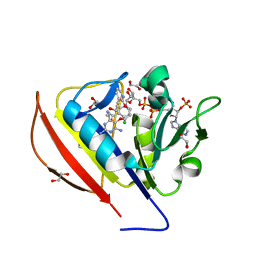
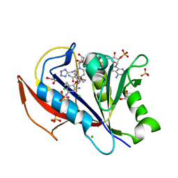
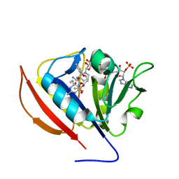
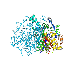
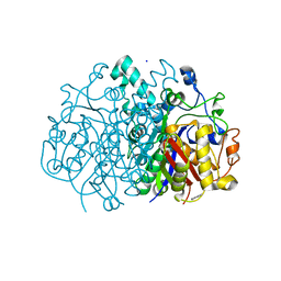
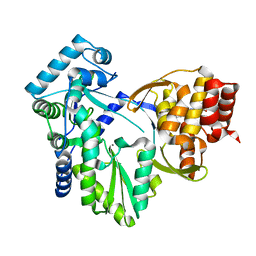
Structure of MK-3281, a Potent Non-Nucleoside Finger-Loop Inhibitor, in complex with the Hepatitis C Virus NS5B Polymerase
Descriptor: (7R)-14-cyclohexyl-7-{[2-(dimethylamino)ethyl](methyl)amino}-7,8-dihydro-6H-indolo[1,2-e][1,5]benzoxazocine-11-carboxylic acid, MANGANESE (II) ION, RNA-DIRECTED RNA POLYMERASE
Authors:Di Marco, S, Baiocco, P.
Deposit date:2010-11-06
Release date:2010-12-22
Last modified:2023-12-20
Method:X-RAY DIFFRACTION (2.53 Å)
Cite:Discovery of (7R)-14-Cyclohexyl-7-{[2-(Dimethylamino)Ethyl] (Methyl)Amino}-7,8-Dihydro-6H-Indolo[1,2-E][1,5] Benzoxazocine -11-Carboxylic Acid (Mk-3281), a Potent and Orally Bioavailable Finger-Loop Inhibitor of the Hepatitis C Virus Ns5B Polymerase
J.Med.Chem., 54, 2011
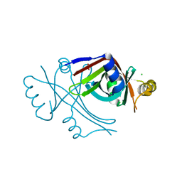
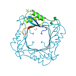
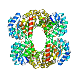
3SI9
DownloadVisualize
BU of 3si9 by Molmil
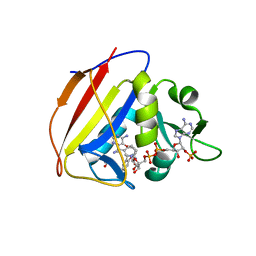
Crystal structure of Dihydrodipicolinate Synthase from Bartonella Henselae
Descriptor: 1,2-ETHANEDIOL, Dihydrodipicolinate synthase
Authors:Seattle Structural Genomics Center for Infectious Disease (SSGCID), Staker, B.L, Abendroth, J, Sankaran, B.
Deposit date:2011-06-17
Release date:2011-06-29
Last modified:2023-09-13
Method:X-RAY DIFFRACTION (2.1 Å)
Cite:Cloning, expression, purification, crystallization and X-ray diffraction analysis of dihydrodipicolinate synthase from the human pathogenic bacterium Bartonella henselae strain Houston-1 at 2.1 angstrom resolution.
Acta Crystallogr F Struct Biol Commun, 72, 2016

224201

건을2024-08-28부터공개중

PDB statisticsPDBj update infoContact PDBjnumon