Loading
PDBj
MenuPDBj@FacebookPDBj@TwitterPDBj@YouTubewwPDB FoundationwwPDB
RCSB PDBPDBeBMRBAdv. SearchSearch help
Search by PDB author
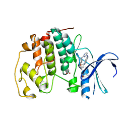
4RJ7
DownloadVisualize
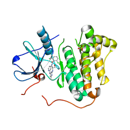
BU of 4rj7 by Molmil
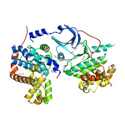
EGFR kinase (T790M/L858R) with inhibitor compound 1
Descriptor: 2,6-dichloro-N-{2-[(2-{[(2S)-1-hydroxypropan-2-yl]amino}-6-methylpyrimidin-4-yl)amino]pyridin-4-yl}benzamide, Epidermal growth factor receptor, SULFATE ION
Authors:Eigenbrot, C, Yu, C.
Deposit date:2014-10-08
Release date:2014-11-26
Last modified:2023-09-20
Method:X-RAY DIFFRACTION (2.55 Å)
Cite:Discovery of Selective and Noncovalent Diaminopyrimidine-Based Inhibitors of Epidermal Growth Factor Receptor Containing the T790M Resistance Mutation.
J.Med.Chem., 57, 2014
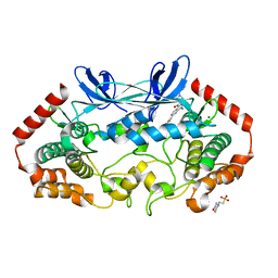
4RJ3
DownloadVisualize
BU of 4rj3 by Molmil
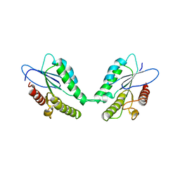
CDK2 with EGFR inhibitor compound 8
Descriptor: 1-cyclopentyl-N-[2-(4-methoxypiperidin-1-yl)pyrimidin-4-yl]-1H-pyrrolo[3,2-c]pyridin-6-amine, ACETATE ION, Cyclin-dependent kinase 2
Authors:Eigenbrot, C, Yin, J.
Deposit date:2014-10-08
Release date:2014-11-26
Last modified:2024-11-06
Method:X-RAY DIFFRACTION (1.63 Å)
Cite:Discovery of Selective and Noncovalent Diaminopyrimidine-Based Inhibitors of Epidermal Growth Factor Receptor Containing the T790M Resistance Mutation.
J.Med.Chem., 57, 2014
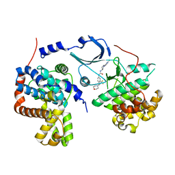
4ZK5
DownloadVisualize
BU of 4zk5 by Molmil
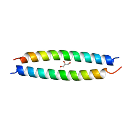
MAP4K4 in complex with inhibitor GNE-495
Descriptor: 2-(N-MORPHOLINO)-ETHANESULFONIC ACID, 8-amino-N-[1-(cyclopropylcarbonyl)azetidin-3-yl]-2-(3-fluorophenyl)-1,7-naphthyridine-5-carboxamide, MAGNESIUM ION, ...
Authors:Harris, S.F, Wu, P, Coons, M.
Deposit date:2015-04-29
Release date:2015-09-02
Last modified:2024-03-06
Method:X-RAY DIFFRACTION (2.89 Å)
Cite:Structure-Based Design of GNE-495, a Potent and Selective MAP4K4 Inhibitor with Efficacy in Retinal Angiogenesis.
Acs Med.Chem.Lett., 6, 2015
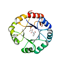
5HVY
DownloadVisualize
BU of 5hvy by Molmil
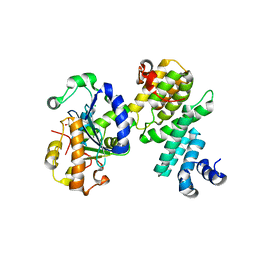
CDK8/CYCC IN COMPLEX WITH COMPOUND 20
Descriptor: CHLORIDE ION, Cyclin-C, Cyclin-dependent kinase 8, ...
Authors:Kiefer, J.R, Schneider, E.V, Maskos, K, Bergeron, P, Koehler, M.
Deposit date:2016-01-28
Release date:2016-04-20
Last modified:2024-03-06
Method:X-RAY DIFFRACTION (2.39 Å)
Cite:Design and Development of a Series of Potent and Selective Type II Inhibitors of CDK8.
Acs Med.Chem.Lett., 7, 2016
1LBM
DownloadVisualize
BU of 1lbm by Molmil
CRYSTAL STRUCTURE OF PHOSPHORIBOSYL ANTHRANILATE ISOMERASE (PRAI) IN COMPLEX WITH REDUCED 1-(O-CARBOXYPHENYLAMINO)-1-DEOXYRIBULOSE 5-PHOSPHATE (RCDRP)
Descriptor: 1-(O-CARBOXY-PHENYLAMINO)-1-DEOXY-D-RIBULOSE-5-PHOSPHATE, PHOSPHORIBOSYL ANTHRANILATE ISOMERASE
Authors:Hennig, M, Sterner, R, Kirschner, K.
Deposit date:2002-04-04
Release date:2002-12-18
Last modified:2023-08-16
Method:X-RAY DIFFRACTION (2.8 Å)
Cite:Two (betaalpha)(8)-barrel enzymes of histidine and tryptophan biosynthesis have similar reaction mechanisms and common strategies for protecting their labile substrates
Biochemistry, 41, 2002
7ZR2
DownloadVisualize
BU of 7zr2 by Molmil
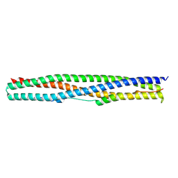
Crystal structure of a chimeric protein mimic of SARS-CoV-2 Spike HR1 in complex with HR2
Descriptor: Spike protein S2', Spike protein S2',Chimeric protein mimic of SARS-CoV-2 Spike HR1
Authors:Camara-Artigas, A, Gavira, J.A, Cano-Munoz, M, Polo-Megias, D, Conejero-Lara, F.
Deposit date:2022-05-03
Release date:2022-11-09
Last modified:2024-10-09
Method:X-RAY DIFFRACTION (1.45 Å)
Cite:Novel chimeric proteins mimicking SARS-CoV-2 spike epitopes with broad inhibitory activity.
Int.J.Biol.Macromol., 222, 2022
2YKO
DownloadVisualize
BU of 2yko by Molmil
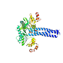
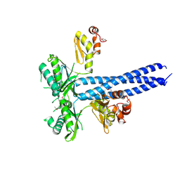
Structure of the human LINE-1 ORF1p trimer
Descriptor: CHLORIDE ION, LINE-1 ORF1P
Authors:Khazina, E, Weichenrieder, O.
Deposit date:2011-05-28
Release date:2011-08-10
Last modified:2023-12-20
Method:X-RAY DIFFRACTION (2.1 Å)
Cite:Trimeric Structure and Flexibility of the L1Orf1P Protein in Human L1 Retrotransposition
Nat.Struct.Mol.Biol., 18, 2011
2YHB
DownloadVisualize
BU of 2yhb by Molmil
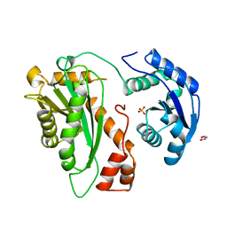
Crystal Structure of the N. crassa QDE-2 AGO MID-PIWI Domains
Descriptor: GLYCEROL, POST-TRANSCRIPTIONAL GENE SILENCING PROTEIN QDE-2, SULFATE ION
Authors:Boland, A, Weichenrieder, O.
Deposit date:2011-04-27
Release date:2011-06-15
Last modified:2023-12-20
Method:X-RAY DIFFRACTION (3.65 Å)
Cite:Crystal Structure of the Mid-Piwi Lobe of a Eukaryotic Argonaute Protein
Proc.Natl.Acad.Sci.USA, 108, 2011
2YKP
DownloadVisualize
BU of 2ykp by Molmil
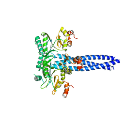
Structure of the human LINE-1 ORF1p trimer
Descriptor: CHLORIDE ION, LINE-1 ORF1P
Authors:Khazina, E, Weichenrieder, O.
Deposit date:2011-05-28
Release date:2011-08-10
Last modified:2023-12-20
Method:X-RAY DIFFRACTION (3.1 Å)
Cite:Trimeric Structure and Flexibility of the L1Orf1P Protein in Human L1 Retrotransposition
Nat.Struct.Mol.Biol., 18, 2011
2YKQ
DownloadVisualize
BU of 2ykq by Molmil
Structure of the human LINE-1 ORF1p trimer
Descriptor: CHLORIDE ION, LINE-1 ORF1P
Authors:Khazina, E, Weichenrieder, O.
Deposit date:2011-05-28
Release date:2011-08-10
Last modified:2024-10-23
Method:X-RAY DIFFRACTION (3.1 Å)
Cite:Trimeric Structure and Flexibility of the L1Orf1P Protein in Human L1 Retrotransposition
Nat.Struct.Mol.Biol., 18, 2011
2YHA
DownloadVisualize
BU of 2yha by Molmil
Crystal Structure of the N. crassa QDE-2 AGO MID-PIWI Domains
Descriptor: GLYCEROL, POST-TRANSCRIPTIONAL GENE SILENCING PROTEIN QDE-2, SULFATE ION
Authors:Boland, A, Weichenrieder, O.
Deposit date:2011-04-27
Release date:2011-06-15
Last modified:2023-12-20
Method:X-RAY DIFFRACTION (1.85 Å)
Cite:Crystal Structure of the Mid-Piwi Lobe of a Eukaryotic Argonaute Protein
Proc.Natl.Acad.Sci.USA, 108, 2011
7BL4
DownloadVisualize
BU of 7bl4 by Molmil
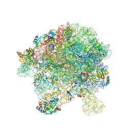
in vitro reconstituted 50S-ObgE-GMPPNP-RsfS particle
Descriptor: 23S ribosomal RNA, 50S ribosomal protein L13, 50S ribosomal protein L14, ...
Authors:Hilal, T, Nikolay, R, Spahn, C.M.T.
Deposit date:2021-01-18
Release date:2021-05-12
Last modified:2024-05-01
Method:ELECTRON MICROSCOPY (2.4 Å)
Cite:Snapshots of native pre-50S ribosomes reveal a biogenesis factor network and evolutionary specialization.
Mol.Cell, 81, 2021
7BL3
DownloadVisualize
BU of 7bl3 by Molmil
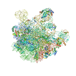
pre-50S-ObgE particle state 2
Descriptor: 23S ribosomal RNA, 50S ribosomal protein L11, 50S ribosomal protein L13, ...
Authors:Hilal, T, Nikolay, R, Spahn, C.M.T.
Deposit date:2021-01-18
Release date:2021-05-12
Last modified:2024-10-23
Method:ELECTRON MICROSCOPY (3.5 Å)
Cite:Snapshots of native pre-50S ribosomes reveal a biogenesis factor network and evolutionary specialization.
Mol.Cell, 81, 2021
5CEI
DownloadVisualize
BU of 5cei by Molmil
Crystal structure of CDK8:Cyclin C complex with compound 22
Descriptor: 1,2-ETHANEDIOL, 4-(4-iodophenoxy)-N-methylthieno[2,3-c]pyridine-2-carboxamide, Cyclin-C, ...
Authors:Kiefer, J.R, Schneider, E.V, Maskos, K, Koehler, M.F.T.
Deposit date:2015-07-06
Release date:2016-02-10
Last modified:2024-03-06
Method:X-RAY DIFFRACTION (2.24 Å)
Cite:Development of a Potent, Specific CDK8 Kinase Inhibitor Which Phenocopies CDK8/19 Knockout Cells.
Acs Med.Chem.Lett., 7, 2016
4C1B
DownloadVisualize
BU of 4c1b by Molmil
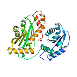
Esterase domain of the ZfL2-1 ORF1 protein from the zebrafish ZfL2-1 retrotransposon
Descriptor: ORF1-ENCODED PROTEIN
Authors:Schneider, A.M, Weichenrieder, O.
Deposit date:2013-08-11
Release date:2013-09-11
Last modified:2024-05-08
Method:X-RAY DIFFRACTION (2.501 Å)
Cite:Structure and Properties of the Esterase from Non-Ltr Retrotransposons Suggest a Role for Lipids in Retrotransposition.
Nucleic Acids Res., 41, 2013
4C1A
DownloadVisualize
BU of 4c1a by Molmil
Coiled coil domain of the ZfL2-1 ORF1 protein from the zebrafish ZfL2- 1 retrotransposon
Descriptor: GLYCEROL, ORF1-ENCODED PROTEIN
Authors:Schneider, A.M, Weichenrieder, O.
Deposit date:2013-08-11
Release date:2013-09-11
Last modified:2023-12-20
Method:X-RAY DIFFRACTION (1.55 Å)
Cite:Structure and Properties of the Esterase from Non-Ltr Retrotransposons Suggest a Role for Lipids in Retrotransposition.
Nucleic Acids Res., 41, 2013
4GMJ
DownloadVisualize
BU of 4gmj by Molmil
Structure of human NOT1 MIF4G domain co-crystallized with CAF1
Descriptor: CCR4-NOT transcription complex subunit 1, CCR4-NOT transcription complex subunit 7, CHLORIDE ION, ...
Authors:Petit, P, Weichenrieder, O, Wohlbold, L, Izaurralde, E.
Deposit date:2012-08-16
Release date:2012-10-03
Last modified:2023-11-08
Method:X-RAY DIFFRACTION (2.7 Å)
Cite:The structural basis for the interaction between the CAF1 nuclease and the NOT1 scaffold of the human CCR4-NOT deadenylase complex
Nucleic Acids Res., 40, 2012
4GML
DownloadVisualize
BU of 4gml by Molmil
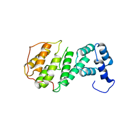
Crystal structure of human NOT1 MIF4G domain
Descriptor: CCR4-NOT transcription complex subunit 1
Authors:Petit, P, Weichenrieder, O, Wohlbold, L, Izaurralde, E.
Deposit date:2012-08-16
Release date:2012-10-03
Last modified:2023-11-08
Method:X-RAY DIFFRACTION (2.9 Å)
Cite:The structural basis for the interaction between the CAF1 nuclease and the NOT1 scaffold of the human CCR4-NOT deadenylase complex
Nucleic Acids Res., 40, 2012
6HQD
DownloadVisualize
BU of 6hqd by Molmil
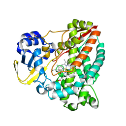
Cytochrome P450-153 from Pseudomonas sp. 19-rlim
Descriptor: Cytochrome P450, GLYCEROL, PROTOPORPHYRIN IX CONTAINING FE, ...
Authors:Fiorentini, F, Mattevi, A.
Deposit date:2018-09-24
Release date:2018-12-12
Last modified:2024-01-24
Method:X-RAY DIFFRACTION (1.8 Å)
Cite:The Extreme Structural Plasticity in the CYP153 Subfamily of P450s Directs Development of Designer Hydroxylases.
Biochemistry, 57, 2018
6HQW
DownloadVisualize
BU of 6hqw by Molmil
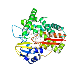
Cytochrome P450-153 from Novosphingobium aromaticivorans
Descriptor: Cytochrome P450, PROTOPORPHYRIN IX CONTAINING FE
Authors:Fiorentini, F, Mattevi, A.
Deposit date:2018-09-25
Release date:2018-12-12
Last modified:2024-01-24
Method:X-RAY DIFFRACTION (2.9 Å)
Cite:The Extreme Structural Plasticity in the CYP153 Subfamily of P450s Directs Development of Designer Hydroxylases.
Biochemistry, 57, 2018
6HQG
DownloadVisualize
BU of 6hqg by Molmil
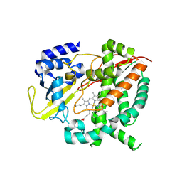
Cytochrome P450-153 from Phenylobacterium zucineum
Descriptor: Cytochrome P450, PROTOPORPHYRIN IX CONTAINING FE
Authors:Fiorentini, F, Mattevi, A.
Deposit date:2018-09-25
Release date:2018-12-12
Last modified:2024-11-06
Method:X-RAY DIFFRACTION (2.9 Å)
Cite:The Extreme Structural Plasticity in the CYP153 Subfamily of P450s Directs Development of Designer Hydroxylases.
Biochemistry, 57, 2018
6T6E
DownloadVisualize
BU of 6t6e by Molmil
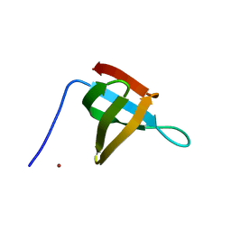
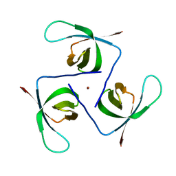
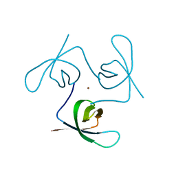
Crystal Structure of the C-terminal domain of the HIV-1 Integrase (PNL4-3)
Descriptor: NICKEL (II) ION, Pol protein
Authors:Ruff, M, Negroni, M.
Deposit date:2019-10-18
Release date:2020-07-22
Last modified:2024-01-24
Method:X-RAY DIFFRACTION (1.302 Å)
Cite:NKNK: a New Essential Motif in the C-Terminal Domain of HIV-1 Group M Integrases.
J.Virol., 94, 2020
6T6J
DownloadVisualize
BU of 6t6j by Molmil
Crystal Structure of the C-terminal domain of the HIV-1 Integrase (subtype A2, mutant N254K, K340Q)
Descriptor: NICKEL (II) ION, Pol protein
Authors:Ruff, M, Negroni, M.
Deposit date:2019-10-18
Release date:2020-07-22
Last modified:2024-01-24
Method:X-RAY DIFFRACTION (2.003 Å)
Cite:NKNK: a New Essential Motif in the C-Terminal Domain of HIV-1 Group M Integrases.
J.Virol., 94, 2020
6T6I
DownloadVisualize
BU of 6t6i by Molmil
Crystal Structure of the C-terminal domain of the HIV-1 Integrase (subtype A2)
Descriptor: NICKEL (II) ION, Pol protein
Authors:Ruff, M, Negroni, M.
Deposit date:2019-10-18
Release date:2020-07-22
Last modified:2024-01-24
Method:X-RAY DIFFRACTION (2.203 Å)
Cite:NKNK: a New Essential Motif in the C-Terminal Domain of HIV-1 Group M Integrases.
J.Virol., 94, 2020
4Q37
DownloadVisualize
BU of 4q37 by Molmil
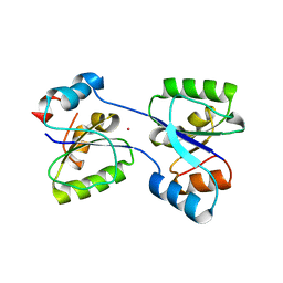
Crystal structure of the hypothetical protein TM0182 Thermotoga maritima, N-terminal domain.
Descriptor: PLATINUM (II) ION, Radical SAM protein
Authors:Hocker, B, Farias-Rico, J.A.
Deposit date:2014-04-11
Release date:2014-07-16
Last modified:2024-02-28
Method:X-RAY DIFFRACTION (3.19 Å)
Cite:Evolutionary relationship of two ancient protein superfolds.
Nat.Chem.Biol., 10, 2014

227111

건을2024-11-06부터공개중

PDB statisticsPDBj update infoContact PDBjnumon