1GRF
 
 | |
1GRA
 | |
1GRE
 | |
2AK2
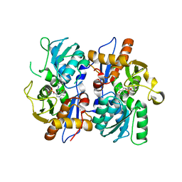
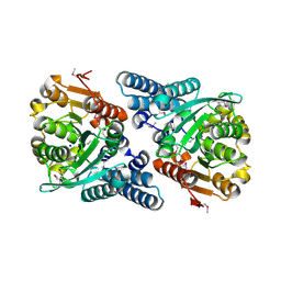
 | | ADENYLATE KINASE ISOENZYME-2 | | Descriptor: | ADENYLATE KINASE ISOENZYME-2, SULFATE ION | | Authors: | Schlauderer, G.J, Schulz, G.E. | | Deposit date: | 1995-12-29 | | Release date: | 1996-06-10 | | Last modified: | 2024-10-30 | | Method: | X-RAY DIFFRACTION (2.1 Å) | | Cite: | The structure of bovine mitochondrial adenylate kinase: comparison with isoenzymes in other compartments.
Protein Sci., 5, 1996
|
|
2BHT
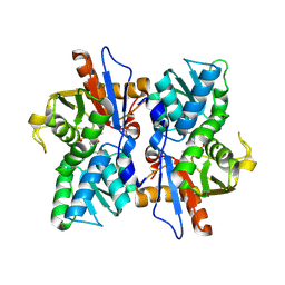
 | | Crystal structure of O-acetylserine sulfhydrylase B | | Descriptor: | CYSTEINE SYNTHASE B, PYRIDOXAL-5'-PHOSPHATE | | Authors: | Claus, M.T, Zocher, G.E, Maier, T.H.P, Schulz, G.E. | | Deposit date: | 2005-01-18 | | Release date: | 2005-06-22 | | Last modified: | 2023-12-13 | | Method: | X-RAY DIFFRACTION (2.1 Å) | | Cite: | Structure of the O-Acetylserine Sulfhydrylase Isoenzyme Cysm from Escherichia Coli
Biochemistry, 44, 2005
|
|
2BHS
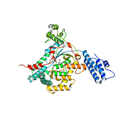
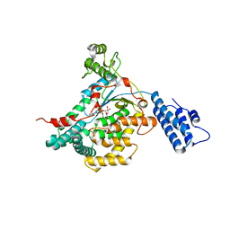
 | | Crystal Structure of Cysteine Synthase B | | Descriptor: | CYSTEINE SYNTHASE B, PYRIDOXAL-5'-PHOSPHATE | | Authors: | Claus, M.T, Zocher, G.E, Maier, T.H.P, Schulz, G.E. | | Deposit date: | 2005-01-18 | | Release date: | 2005-06-22 | | Last modified: | 2025-04-09 | | Method: | X-RAY DIFFRACTION (2.67 Å) | | Cite: | Structure of the O-Acetylserine Sulfhydrylase Isoenzyme Cysm from Escherichia Coli
Biochemistry, 44, 2005
|
|
2VL8
 | | CRYSTAL STRUCTURE OF THE CATALYTIC DOMAIN OF LETHAL TOXIN FROM CLOSTRIDIUM SORDELLII IN COMPLEX WITH UDP, CASTANOSPERMINE AND CALCIUM ION | | Descriptor: | CALCIUM ION, CASTANOSPERMINE, CYTOTOXIN L, ... | | Authors: | Jank, T, Ziegler, M.O.P, Schulz, G.E, Aktories, K. | | Deposit date: | 2008-01-09 | | Release date: | 2008-06-17 | | Last modified: | 2024-05-08 | | Method: | X-RAY DIFFRACTION (2.31 Å) | | Cite: | Inhibition of the Glucosyltransferase Activity of Clostridial Rho/Ras-Glucosylating Toxins by Castanospermine.
FEBS Lett., 582, 2008
|
|
2VKH
 | | CRYSTAL STRUCTURE OF THE CATALYTIC DOMAIN OF LETHAL TOXIN FROM CLOSTRIDIUM SORDELLII IN COMPLEX WITH UDP-GLC AND CALCIUM ION | | Descriptor: | CALCIUM ION, CYTOTOXIN L, URIDINE-5'-DIPHOSPHATE-GLUCOSE | | Authors: | Ziegler, M.O.P, Jank, T, Aktories, K, Schulz, G.E. | | Deposit date: | 2007-12-19 | | Release date: | 2008-03-18 | | Last modified: | 2024-05-08 | | Method: | X-RAY DIFFRACTION (2.3 Å) | | Cite: | Conformational Changes and Reaction of Clostridial Glycosylating Toxins.
J.Mol.Biol., 377, 2008
|
|
2VK9
 | |
2VQL
 | |
2VQG
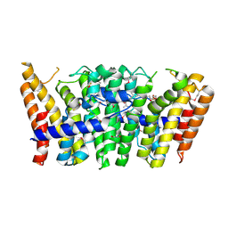
 | | Crystal structure of PorB from Corynebacterium glutamicum (crystal form I) | | Descriptor: | (4R)-2-METHYLPENTANE-2,4-DIOL, 3,6,9,12,15,18,21,24,27-nonaoxaheptatriacontan-1-ol, ACETATE ION, ... | | Authors: | Ziegler, K, Benz, R, Schulz, G.E. | | Deposit date: | 2008-03-15 | | Release date: | 2008-05-20 | | Last modified: | 2024-10-16 | | Method: | X-RAY DIFFRACTION (1.82 Å) | | Cite: | A Putative Alpha-Helical Porin from Corynebacterium Glutamicum.
J.Mol.Biol., 379, 2008
|
|
2POR
 | |
2VQH
 | |
2VSE
 | | Structure and mode of action of a mosquitocidal holotoxin | | Descriptor: | (4S)-2-METHYL-2,4-PENTANEDIOL, GLYCEROL, MOSQUITOCIDAL TOXIN | | Authors: | Treiber, N, Reinert, D.J, Carpusca, I, Aktories, K, Schulz, G.E. | | Deposit date: | 2008-04-22 | | Release date: | 2008-07-08 | | Last modified: | 2023-12-13 | | Method: | X-RAY DIFFRACTION (2.5 Å) | | Cite: | Structure and Mode of Action of a Mosquitocidal Holotoxin.
J.Mol.Biol., 381, 2008
|
|
2VOU
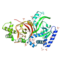
 | | Structure of 2,6-dihydroxypyridine-3-hydroxylase from Arthrobacter nicotinovorans | | Descriptor: | 2,6-DIHYDROXYPYRIDINE HYDROXYLASE, ACETATE ION, FLAVIN-ADENINE DINUCLEOTIDE, ... | | Authors: | Treiber, N, Schulz, G.E. | | Deposit date: | 2008-02-21 | | Release date: | 2008-05-13 | | Last modified: | 2024-05-08 | | Method: | X-RAY DIFFRACTION (2.6 Å) | | Cite: | Structure of 2,6-Dihydroxypyridine 3-Hydroxylase from a Nicotine-Degrading Pathway.
J.Mol.Biol., 379, 2008
|
|
2VQK
 | |
2VSA
 | | Structure and mode of action of a mosquitocidal holotoxin | | Descriptor: | MOSQUITOCIDAL TOXIN | | Authors: | Treiber, N, Reinert, D.J, Carpusca, I, Aktories, K, Schulz, G.E. | | Deposit date: | 2008-04-22 | | Release date: | 2008-07-08 | | Last modified: | 2023-12-13 | | Method: | X-RAY DIFFRACTION (2.89 Å) | | Cite: | Structure and Mode of Action of a Mosquitocidal Holotoxin
J.Mol.Biol., 381, 2008
|
|
2J42
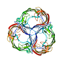
 | | low quality crystal structure of the transport component C2-II of the C2-toxin from Clostridium botulinum | | Descriptor: | C2 TOXIN COMPONENT-II | | Authors: | Schleberger, C, Hochmann, H, Barth, H, Aktories, K, Schulz, G.E. | | Deposit date: | 2006-08-24 | | Release date: | 2006-10-11 | | Last modified: | 2023-12-13 | | Method: | X-RAY DIFFRACTION (3.13 Å) | | Cite: | Structure and Action of the Binary C2 Toxin from Clostridium Botulinum.
J.Mol.Biol., 364, 2006
|
|
2J9A
 | | blLAP in Complex with Microginin FR1 | | Descriptor: | (2S,3R)-3-AMINO-2-HYDROXYDECANOIC ACID, (4S)-2-METHYL-2,4-PENTANEDIOL, CHLORIDE ION, ... | | Authors: | Kraft, M, Schleberger, C, Weckesser, J, Schulz, G.E. | | Deposit date: | 2006-11-06 | | Release date: | 2006-12-06 | | Last modified: | 2024-11-06 | | Method: | X-RAY DIFFRACTION (1.73 Å) | | Cite: | Binding Structure of the Leucine Aminopeptidase Inhibitor Microginin Fr1.
FEBS Lett., 580, 2006
|
|
2J3V
 | | Crystal structure of the enzymatic component C2-I of the C2-toxin from Clostridium botulinum at pH 3.0 | | Descriptor: | C2 TOXIN COMPONENT I, GLYCEROL, SULFATE ION | | Authors: | Schleberger, C, Hochmann, H, Barth, H, Aktories, K, Schulz, G.E. | | Deposit date: | 2006-08-23 | | Release date: | 2006-10-11 | | Last modified: | 2024-05-08 | | Method: | X-RAY DIFFRACTION (2.11 Å) | | Cite: | Structure and Action of the Binary C2 Toxin from Clostridium Botulinum.
J.Mol.Biol., 364, 2006
|
|
2J1P
 | | Geranylgeranyl diphosphate synthase from Sinapis alba in complex with GGPP | | Descriptor: | BETA-MERCAPTOETHANOL, GERANYLGERANYL DIPHOSPHATE, GERANYLGERANYL PYROPHOSPHATE SYNTHETASE, ... | | Authors: | Kloer, D.P, Welsch, R, Beyer, P, Schulz, G.E. | | Deposit date: | 2006-08-15 | | Release date: | 2007-01-02 | | Last modified: | 2025-04-09 | | Method: | X-RAY DIFFRACTION (1.8 Å) | | Cite: | Structure and Reaction Geometry of Geranylgeranyl Diphosphate Synthase from Sinapis Alba.
Biochemistry, 45, 2006
|
|
2J3X
 | | Crystal structure of the enzymatic component C2-I of the C2-toxin from Clostridium botulinum at pH 3.0 (mut-S361R) | | Descriptor: | C2 TOXIN COMPONENT I, GLYCEROL, SULFATE ION | | Authors: | Schleberger, C, Hochmann, H, Barth, H, Aktories, K, Schulz, G.E. | | Deposit date: | 2006-08-23 | | Release date: | 2006-10-11 | | Last modified: | 2023-12-13 | | Method: | X-RAY DIFFRACTION (1.75 Å) | | Cite: | Structure and Action of the Binary C2 Toxin from Clostridium Botulinum.
J.Mol.Biol., 364, 2006
|
|
2JBW
 | | Crystal Structure of the 2,6-dihydroxy-pseudo-oxynicotine Hydrolase. | | Descriptor: | 2,6-DIHYDROXY-PSEUDO-OXYNICOTINE HYDROLASE, SODIUM ION | | Authors: | Schleberger, C, Sachelaru, P, Brandsch, R, Schulz, G.E. | | Deposit date: | 2006-12-14 | | Release date: | 2007-01-04 | | Last modified: | 2024-11-06 | | Method: | X-RAY DIFFRACTION (2.1 Å) | | Cite: | Structure and Action of a Cc Bond Cleaving Alpha/Beta-Hydrolase Involved in Nicotine Degration.
J.Mol.Biol., 367, 2007
|
|
2MPR
 | | MALTOPORIN FROM SALMONELLA TYPHIMURIUM | | Descriptor: | CALCIUM ION, MALTOPORIN, alpha-D-glucopyranose-(1-4)-alpha-D-glucopyranose-(1-4)-beta-D-glucopyranose | | Authors: | Meyer, J.E.W, Schulz, G.E. | | Deposit date: | 1997-02-07 | | Release date: | 1997-04-01 | | Last modified: | 2024-10-23 | | Method: | X-RAY DIFFRACTION (2.4 Å) | | Cite: | Structure of maltoporin from Salmonella typhimurium ligated with a nitrophenyl-maltotrioside.
J.Mol.Biol., 266, 1997
|
|
3E1T
 | | Structure and action of the myxobacterial chondrochloren halogenase CndH, a new variant of FAD-dependent halogenases | | Descriptor: | CHLORIDE ION, FLAVIN-ADENINE DINUCLEOTIDE, Halogenase | | Authors: | Buedenbender, S, Rachid, S, Mueller, R, Schulz, G.E. | | Deposit date: | 2008-08-04 | | Release date: | 2009-02-24 | | Last modified: | 2024-03-20 | | Method: | X-RAY DIFFRACTION (2.05 Å) | | Cite: | Structure and action of the myxobacterial chondrochloren halogenase CndH: a new variant of FAD-dependent halogenases.
J.Mol.Biol., 385, 2009
|
|


