Loading
PDBj
MenuPDBj@FacebookPDBj@TwitterPDBj@YouTubewwPDB FoundationwwPDB
RCSB PDBPDBeBMRBAdv. SearchSearch help
Search by PDB author
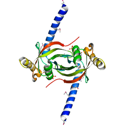
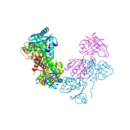
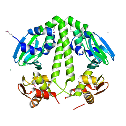
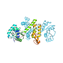
1VL0
DownloadVisualize
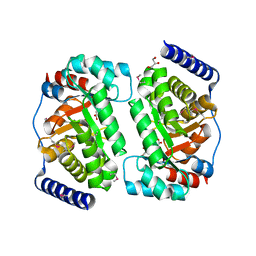
BU of 1vl0 by Molmil
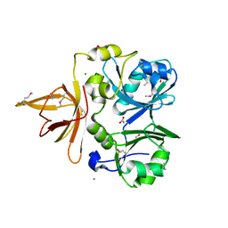
CRYSTAL STRUCTURE OF A DTDP-4-DEHYDRORHAMNOSE REDUCTASE, RFBD ORTHOLOG (CA_C2315) FROM CLOSTRIDIUM ACETOBUTYLICUM ATCC 824 AT 2.05 A RESOLUTION
Descriptor: 1,4-DIHYDRONICOTINAMIDE ADENINE DINUCLEOTIDE, DTDP-4-dehydrorhamnose reductase, rfbD ortholog, ...
Authors:Joint Center for Structural Genomics (JCSG)
Deposit date:2004-06-30
Release date:2004-08-17
Last modified:2024-10-30
Method:X-RAY DIFFRACTION (2.05 Å)
Cite:Crystal structure of DTDP-4-dehydrorhamnose reductase, rfbD ortholog (CAC2315) from Clostridium acetobutylicum at 2.05 A resolution
To be published
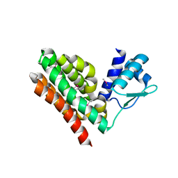
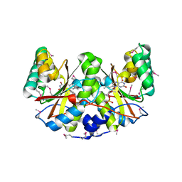
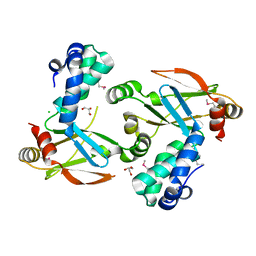
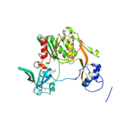
1VLL
DownloadVisualize
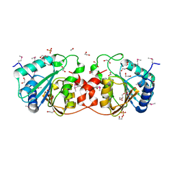
BU of 1vll by Molmil
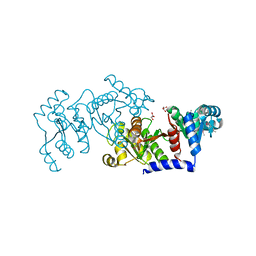
Crystal structure of alanine dehydrogenase (AF1665) from Archaeoglobus fulgidus at 2.80 A resolution
Descriptor: alanine dehydrogenase
Authors:Joint Center for Structural Genomics (JCSG)
Deposit date:2004-08-03
Release date:2004-09-21
Last modified:2024-05-22
Method:X-RAY DIFFRACTION (2.8 Å)
Cite:Crystal structure of alanine dehydrogenase (AF1665) from Archaeoglobus fulgidus at 2.80 A resolution
To be published
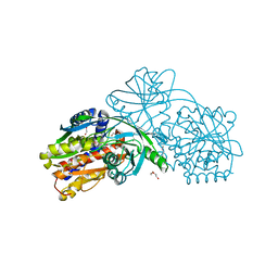
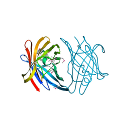
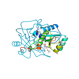
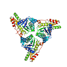
1VLY
DownloadVisualize
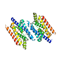
BU of 1vly by Molmil
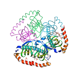
Crystal structure of a putative aminomethyltransferase (ygfz) from escherichia coli at 1.30 A resolution
Descriptor: 1,2-ETHANEDIOL, ACETATE ION, CALCIUM ION, ...
Authors:Joint Center for Structural Genomics (JCSG)
Deposit date:2004-08-20
Release date:2004-08-31
Last modified:2024-10-09
Method:X-RAY DIFFRACTION (1.3 Å)
Cite:Crystal structure of Unknown protein from 2D-page (Spot PR51) (b2898) from Escherichia coli k12 at 1.30 A resolution
To be published
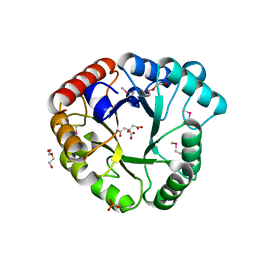
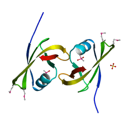
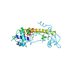
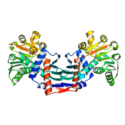
1VMI
DownloadVisualize
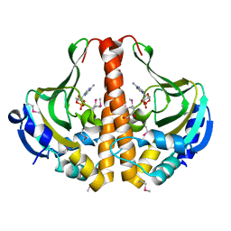
BU of 1vmi by Molmil
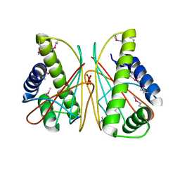
Crystal structure of Putative phosphate acetyltransferase (np_416953.1) from Escherichia coli k12 at 2.32 A resolution
Descriptor: GLYCEROL, putative phosphate acetyltransferase
Authors:Joint Center for Structural Genomics (JCSG)
Deposit date:2004-09-28
Release date:2004-10-12
Last modified:2023-12-27
Method:X-RAY DIFFRACTION (2.32 Å)
Cite:Crystal structure of Putative phosphate acetyltransferase (np_416953.1) from Escherichia coli k12 at 2.32 A resolution
To be published
1VPM
DownloadVisualize
BU of 1vpm by Molmil
Crystal structure of Acyl-CoA hydrolase (NP_241664.1) from Bacillus halodurans at 1.66 A resolution
Descriptor: COENZYME A, PHOSPHATE ION, acyl-CoA hydrolase
Authors:Joint Center for Structural Genomics (JCSG)
Deposit date:2004-11-11
Release date:2004-12-14
Last modified:2023-01-25
Method:X-RAY DIFFRACTION (1.66 Å)
Cite:Crystal structure of Acyl-CoA hydrolase (NP_241664.1) from Bacillus halodurans at 1.66 A resolution
To be published
3HM4
DownloadVisualize
BU of 3hm4 by Molmil
CRYSTAL STRUCTURE OF A CHEMOTAXIS PROTEIN CHEX (DDE_0281) FROM DESULFOVIBRIO DESULFURICANS SUBSP. AT 1.30 A RESOLUTION
Descriptor: Chemotaxis protein CheX, GLYCEROL, UNKNOWN LIGAND
Authors:Joint Center for Structural Genomics (JCSG)
Deposit date:2009-05-28
Release date:2009-06-23
Last modified:2023-02-01
Method:X-RAY DIFFRACTION (1.3 Å)
Cite:Crystal structure of Chemotaxis protein CheX (YP_386777.1) from DESULFOVIBRIO DESULFURICANS G20 at 1.30 A resolution
To be published
3IN6
DownloadVisualize
BU of 3in6 by Molmil
Crystal structure of a fmn-binding protein (swol_0183) from syntrophomonas wolfei subsp. wolfei at 2.12 A resolution
Descriptor: (4S)-2-METHYL-2,4-PENTANEDIOL, FLAVIN MONONUCLEOTIDE, FMN-binding protein
Authors:Joint Center for Structural Genomics (JCSG)
Deposit date:2009-08-11
Release date:2009-08-25
Last modified:2023-02-01
Method:X-RAY DIFFRACTION (2.12 Å)
Cite:Crystal structure of FMN-binding protein (YP_752906.1) from Syntrophomonas wolfei str. Goettingen at 2.12 A resolution
To be published
3IHU
DownloadVisualize
BU of 3ihu by Molmil
Crystal structure of DNA binding protein (YP_298823.1) from Ralstonia eutropha JMP134 at 1.92 A resolution
Descriptor: CHLORIDE ION, GLYCEROL, Transcriptional regulator, ...
Authors:Joint Center for Structural Genomics (JCSG)
Deposit date:2009-07-30
Release date:2009-08-11
Last modified:2023-02-01
Method:X-RAY DIFFRACTION (1.92 Å)
Cite:Crystal structure of DNA binding protein (YP_298823.1) from Ralstonia eutropha JMP134 at 1.92 A resolution
To be published
3IIB
DownloadVisualize
BU of 3iib by Molmil
Crystal structure of Peptidase M28 precursor (YP_926796.1) from SHEWANELLA AMAZONENSIS SB2B at 1.70 A resolution
Descriptor: DI(HYDROXYETHYL)ETHER, Peptidase M28, TRIETHYLENE GLYCOL, ...
Authors:Joint Center for Structural Genomics (JCSG)
Deposit date:2009-07-31
Release date:2009-08-25
Last modified:2024-10-30
Method:X-RAY DIFFRACTION (1.7 Å)
Cite:Crystal structure of Peptidase M28 precursor (YP_926796.1) from SHEWANELLA AMAZONENSIS SB2B at 1.70 A resolution
To be published
3ORU
DownloadVisualize
BU of 3oru by Molmil
Crystal structure of a DUF1989 family protein (TM1040_0329) from SILICIBACTER SP. TM1040 at 1.11 A resolution
Descriptor: CHLORIDE ION, DUF1989 family protein, MAGNESIUM ION, ...
Authors:Joint Center for Structural Genomics (JCSG)
Deposit date:2010-09-07
Release date:2010-10-13
Last modified:2023-02-01
Method:X-RAY DIFFRACTION (1.11 Å)
Cite:Crystal structure of a DUF1989 family protein (TM1040_0329) from SILICIBACTER SP. TM1040 at 1.11 A resolution
To be published
3P6L
DownloadVisualize
BU of 3p6l by Molmil
Crystal structure of a Sugar phosphate isomerase/epimerase (BDI_1903) from Parabacteroides distasonis ATCC 8503 at 1.85 A resolution
Descriptor: CITRIC ACID, DI(HYDROXYETHYL)ETHER, PHOSPHATE ION, ...
Authors:Joint Center for Structural Genomics (JCSG)
Deposit date:2010-10-11
Release date:2010-12-08
Last modified:2023-02-01
Method:X-RAY DIFFRACTION (1.85 Å)
Cite:Crystal structure of a Sugar phosphate isomerase/epimerase (BDI_1903) from Parabacteroides distasonis ATCC 8503 at 1.85 A resolution
To be published
3ON5
DownloadVisualize
BU of 3on5 by Molmil
Crystal structure of a xanthine dehydrogenase (BH1974) from Bacillus halodurans at 2.80 A resolution
Descriptor: BH1974 protein, CHLORIDE ION
Authors:Joint Center for Structural Genomics (JCSG)
Deposit date:2010-08-27
Release date:2010-09-15
Last modified:2023-12-06
Method:X-RAY DIFFRACTION (2.8 Å)
Cite:Crystal structure of a xanthine dehydrogenase (BH1974) from Bacillus halodurans at 2.80 A resolution
To be published
3PXV
DownloadVisualize
BU of 3pxv by Molmil
Crystal structure of a Nitroreductase with bound FMN (Dhaf_2018) from Desulfitobacterium hafniense DCB-2 at 2.30 A resolution
Descriptor: FLAVIN MONONUCLEOTIDE, Nitroreductase
Authors:Joint Center for Structural Genomics (JCSG)
Deposit date:2010-12-10
Release date:2010-12-29
Last modified:2023-02-01
Method:X-RAY DIFFRACTION (2.3 Å)
Cite:Crystal structure of a Nitroreductase with bound FMN (Dhaf_2018) from Desulfitobacterium hafniense DCB-2 at 2.30 A resolution
To be published
3FZX
DownloadVisualize
BU of 3fzx by Molmil
CRYSTAL STRUCTURE OF A PUTATIVE EXPORTED PROTEIN WITH YMCC-LIKE FOLD (BF2203) FROM BACTEROIDES FRAGILIS NCTC 9343 AT 2.22 A RESOLUTION
Descriptor: CALCIUM ION, Putative exported protein
Authors:Joint Center for Structural Genomics (JCSG)
Deposit date:2009-01-26
Release date:2009-02-03
Last modified:2023-02-01
Method:X-RAY DIFFRACTION (2.22 Å)
Cite:Crystal structure of putative exported protein with YmcC-like fold (YP_211880.1) from Bacteroides fragilis NCTC 9343 at 2.22 A resolution
To be published
3IUW
DownloadVisualize
BU of 3iuw by Molmil
Crystal structure of Activating signal cointegrator (NP_814290.1) from ENTEROCOCCUS FAECALIS V583 at 1.58 A resolution
Descriptor: Activating signal cointegrator, CACODYLATE ION, SULFATE ION
Authors:Joint Center for Structural Genomics (JCSG)
Deposit date:2009-08-31
Release date:2009-09-15
Last modified:2023-02-01
Method:X-RAY DIFFRACTION (1.58 Å)
Cite:Crystal structure of Activating signal cointegrator (NP_814290.1) from ENTEROCOCCUS FAECALIS V583 at 1.58 A resolution
To be published
3R12
DownloadVisualize
BU of 3r12 by Molmil
Crystal structure of a Deoxyribose-phosphate aldolase (TM_1559) from THERMOTOGA MARITIMA at 1.75 A resolution
Descriptor: CITRIC ACID, Deoxyribose-phosphate aldolase, GLYCEROL, ...
Authors:Joint Center for Structural Genomics (JCSG)
Deposit date:2011-03-09
Release date:2011-04-20
Last modified:2023-02-01
Method:X-RAY DIFFRACTION (1.75 Å)
Cite:Crystal structure of a Deoxyribose-phosphate aldolase (TM_1559) from THERMOTOGA MARITIMA at 1.75 A resolution
To be published
3CVO
DownloadVisualize
BU of 3cvo by Molmil
Crystal structure of a methyltransferase-like protein (spo2022) from silicibacter pomeroyi dss-3 at 1.80 A resolution
Descriptor: 1,2-ETHANEDIOL, GLYCEROL, Methyltransferase-like protein of unknown function, ...
Authors:Joint Center for Structural Genomics (JCSG)
Deposit date:2008-04-18
Release date:2008-05-06
Last modified:2023-02-01
Method:X-RAY DIFFRACTION (1.8 Å)
Cite:Crystal structure of methyltransferase-like protein of unknown function (YP_167254.1) from Silicibacter pomeroyi DSS-3 at 1.80 A resolution
To be published
3EZ0
DownloadVisualize
BU of 3ez0 by Molmil
Crystal structure of protein of unknown function with ferritin-like fold (YP_832262.1) from Arthrobacter sp. FB24 at 2.33 A resolution
Descriptor: 1,2-ETHANEDIOL, L(+)-TARTARIC ACID, UNKNOWN LIGAND, ...
Authors:Joint Center for Structural Genomics (JCSG)
Deposit date:2008-10-22
Release date:2008-11-25
Last modified:2023-02-01
Method:X-RAY DIFFRACTION (2.33 Å)
Cite:Crystal structure of protein of unknown function with ferritin-like fold (YP_832262.1) from Arthrobacter sp. FB24 at 2.33 A resolution
To be published
3GYD
DownloadVisualize
BU of 3gyd by Molmil
Crystal structure of a cyclic nucleotide-binding domain (mfla_1926) from methylobacillus flagellatus kt at 1.79 A resolution
Descriptor: ADENOSINE-3',5'-CYCLIC-MONOPHOSPHATE, Cyclic nucleotide-binding domain, GLYCEROL
Authors:Joint Center for Structural Genomics (JCSG)
Deposit date:2009-04-03
Release date:2009-04-21
Last modified:2023-02-01
Method:X-RAY DIFFRACTION (1.79 Å)
Cite:Crystal structure of cyclic nucleotide-binding domain (YP_546034.1) from Methylobacillus flagellatus KT at 1.79 A resolution
To be published
3H3Z
DownloadVisualize
BU of 3h3z by Molmil
Crystal structure of a putative cyclic nucleotide binding protein (spoa0323) from ruegeria pomeroyi dss-3 at 2.35 A resolution
Descriptor: CHLORIDE ION, Cyclic nucleotide-binding protein
Authors:Joint Center for Structural Genomics (JCSG)
Deposit date:2009-04-17
Release date:2009-05-05
Last modified:2023-02-01
Method:X-RAY DIFFRACTION (2.35 Å)
Cite:Crystal structure of PUTATIVE CAMP-BINDING REGULATORY PROTEIN (YP_165150.1) from SILICIBACTER POMEROYI DSS-3 at 2.35 A resolution
To be published
3FJV
DownloadVisualize
BU of 3fjv by Molmil
Crystal structure of novel protein of unknown function (YP_111841.1) from BURKHOLDERIA PSEUDOMALLEI K96243 at 1.90 A resolution
Descriptor: CHLORIDE ION, GLYCEROL, uncharacterized novel protein
Authors:Joint Center for Structural Genomics (JCSG)
Deposit date:2008-12-15
Release date:2009-01-13
Last modified:2024-10-30
Method:X-RAY DIFFRACTION (1.9 Å)
Cite:Crystal structure of novel protein of unknown function (YP_111841.1) from BURKHOLDERIA PSEUDOMALLEI K96243 at 1.90 A resolution
To be published
3H4O
DownloadVisualize
BU of 3h4o by Molmil
Crystal structure of a nitroreductase family protein (cd3355) from clostridium difficile 630 at 1.50 A resolution
Descriptor: (4R)-2-METHYLPENTANE-2,4-DIOL, (4S)-2-METHYL-2,4-PENTANEDIOL, FLAVIN MONONUCLEOTIDE, ...
Authors:Joint Center for Structural Genomics (JCSG)
Deposit date:2009-04-20
Release date:2009-05-19
Last modified:2023-02-01
Method:X-RAY DIFFRACTION (1.5 Å)
Cite:Crystal structure of NITROREDUCTASE FAMILY PROTEIN (YP_001089872.1) from CLOSTRIDIUM DIFFICILE 630 at 1.50 A resolution
To be published
3K69
DownloadVisualize
BU of 3k69 by Molmil
CRYSTAL STRUCTURE OF A PUTATIVE TRANSCRIPTIONAL REGULATOR (LP_0360) FROM LACTOBACILLUS PLANTARUM AT 1.95 A RESOLUTION
Descriptor: DIMETHYL SULFOXIDE, Putative transcription regulator
Authors:Joint Center for Structural Genomics (JCSG)
Deposit date:2009-10-08
Release date:2009-11-03
Last modified:2023-02-01
Method:X-RAY DIFFRACTION (1.95 Å)
Cite:Crystal structure of Putative transcriptional regulator (NP_784167.1) from LACTOBACILLUS PLANTARUM at 1.95 A resolution
To be published
1ZEJ
DownloadVisualize
BU of 1zej by Molmil
Crystal structure of the 3-hydroxyacyl-coa dehydrogenase (hbd-9, af2017) from archaeoglobus fulgidus dsm 4304 at 2.00 A resolution
Descriptor: 3,6,9,12,15,18,21-HEPTAOXATRICOSANE-1,23-DIOL, 3-hydroxyacyl-CoA dehydrogenase, CHLORIDE ION
Authors:Joint Center for Structural Genomics (JCSG)
Deposit date:2005-04-18
Release date:2005-05-03
Last modified:2023-01-25
Method:X-RAY DIFFRACTION (2 Å)
Cite:Crystal structure of 3-hydroxyacyl-CoA dehydrogenase (HBD-9) (np_070841.1) from Archaeoglobus fulgidus at 2.00 A resolution
To be published
3K50
DownloadVisualize
BU of 3k50 by Molmil
Crystal structure of Putative S41 protease (YP_211611.1) from Bacteroides fragilis NCTC 9343 at 2.00 A resolution
Descriptor: CHLORIDE ION, GLYCEROL, Putative S41 protease
Authors:Joint Center for Structural Genomics (JCSG)
Deposit date:2009-10-06
Release date:2009-10-13
Last modified:2023-02-01
Method:X-RAY DIFFRACTION (2 Å)
Cite:Crystal structure of Putative S41 protease (YP_211611.1) from Bacteroides fragilis NCTC 9343 at 2.00 A resolution
To be published

226707

건을2024-10-30부터공개중

PDB statisticsPDBj update infoContact PDBjnumon