7DFK
 
 | |
7DTF
 | |
7DTB
 | |
7E02
 | |
7E03
 | |
7E25
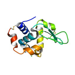
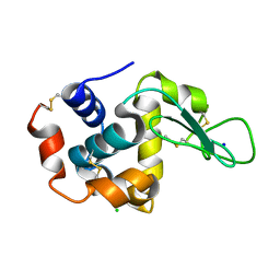
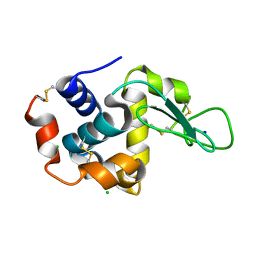
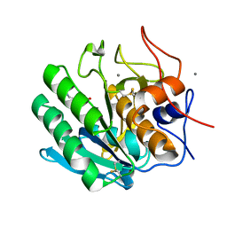
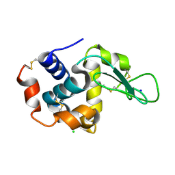
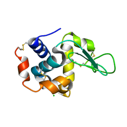
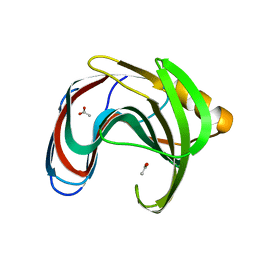
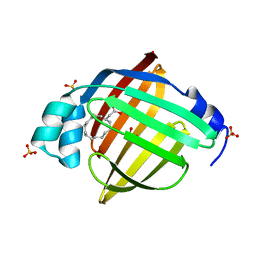 | | Crystal structure of human FABP7 complexed with palmitic acid | | Descriptor: | Fatty acid-binding protein, brain, GLYCEROL, ... | | Authors: | Nam, K.H. | | Deposit date: | 2021-02-04 | | Release date: | 2021-07-14 | | Last modified: | 2023-11-29 | | Method: | X-RAY DIFFRACTION (1.6 Å) | | Cite: | Crystal structure of human brain-type fatty acid-binding protein FABP7 complexed with palmitic acid.
Acta Crystallogr D Struct Biol, 77, 2021
|
|
6K1W
 | |
6KCC
 | |
6K1Y
 | |
6KCB
 | |
6KD2
 | |
6KCD
 | |
6KKF
 | |
6K1X
 | |
6KCA
 | |
6KD1
 | |
6LZO
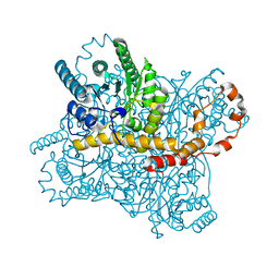
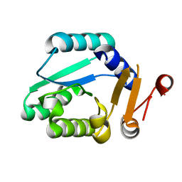
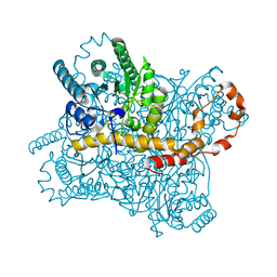
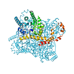
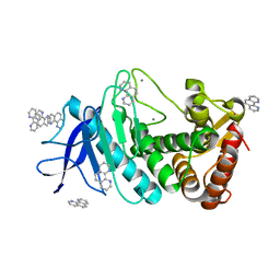 | | Thermolysin with 1,10-phenanthroline | | Descriptor: | 1,10-PHENANTHROLINE, CALCIUM ION, Thermolysin | | Authors: | Nam, K.H. | | Deposit date: | 2020-02-19 | | Release date: | 2021-01-27 | | Last modified: | 2023-11-29 | | Method: | X-RAY DIFFRACTION (1.8 Å) | | Cite: | Structural analysis of metal chelation of the metalloproteinase thermolysin by 1,10-phenanthroline.
J.Inorg.Biochem., 215, 2021
|
|
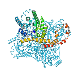
6LZN
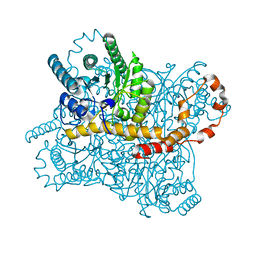
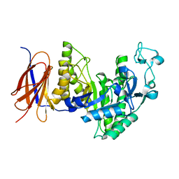
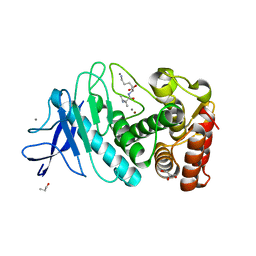 | | Thermolysin | | Descriptor: | CALCIUM ION, GLYCEROL, ISOLEUCINE, ... | | Authors: | Nam, K.H. | | Deposit date: | 2020-02-19 | | Release date: | 2021-01-27 | | Last modified: | 2023-11-29 | | Method: | X-RAY DIFFRACTION (1.5 Å) | | Cite: | Structural analysis of metal chelation of the metalloproteinase thermolysin by 1,10-phenanthroline.
J.Inorg.Biochem., 215, 2021
|
|
6LL2
 | |
6LL3
 | |
6IRK
 | |
6IG6
 | |
3II1
 | |
8IH1
 | |
8IH0
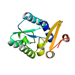
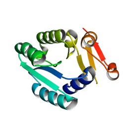
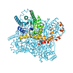
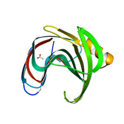
 | | Crystal structure of GH11 from Thermoanaerobacterium saccharolyticum | | Descriptor: | ACETATE ION, Endo-1,4-beta-xylanase | | Authors: | Nam, K.H. | | Deposit date: | 2023-02-22 | | Release date: | 2023-11-22 | | Method: | X-RAY DIFFRACTION (1.5 Å) | | Cite: | Characterization and structural analysis of the endo-1,4-beta-xylanase GH11 from the hemicellulose-degrading Thermoanaerobacterium saccharolyticum useful for lignocellulose saccharification.
Sci Rep, 13, 2023
|
|


