Loading
PDBj
MenuPDBj@FacebookPDBj@TwitterPDBj@YouTubewwPDB FoundationwwPDB
RCSB PDBPDBeBMRBAdv. SearchSearch help
Search by PDB author
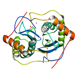
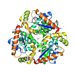
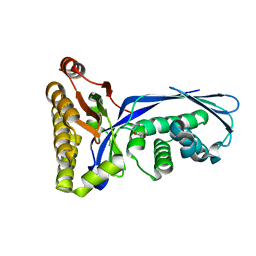
2XWX
DownloadVisualize
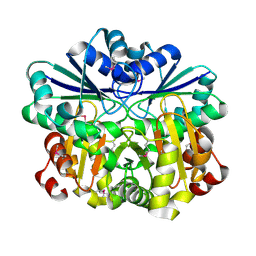
BU of 2xwx by Molmil
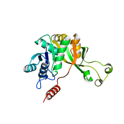
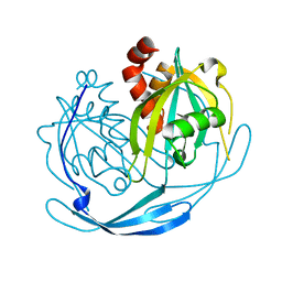
Vibrio cholerae colonization factor GbpA crystal structure
Descriptor: GLCNAC-BINDING PROTEIN A
Authors:Wong, E, Vaaje-Kolstad, G, Ghosh, A, Guerrero, R.H, Konarev, P.V, Ibrahim, A.F.M, Svergun, D.I, Eijsink, V.G.H, Chatterjee, N.S, van Aalten, D.M.F.
Deposit date:2010-11-06
Release date:2011-11-16
Last modified:2015-04-15
Method:X-RAY DIFFRACTION (1.8 Å)
Cite:The Vibrio Cholerae Colonization Factor Gbpa Possesses a Modular Structure that Governs Binding to Different Host Surfaces.
Plos Pathog., 8, 2012
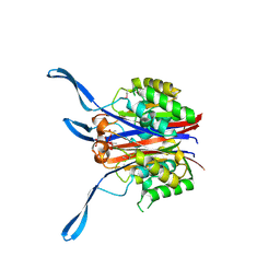
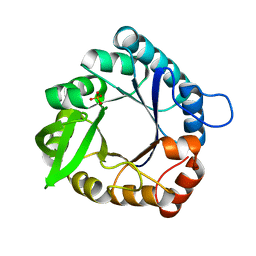
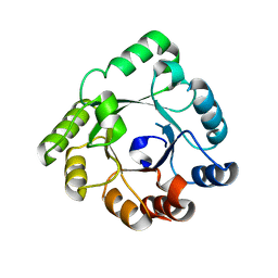
3Q4F
DownloadVisualize
BU of 3q4f by Molmil
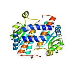
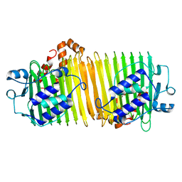
Crystal structure of xrcc4/xlf-cernunnos complex
Descriptor: DNA repair protein XRCC4, Non-homologous end-joining factor 1
Authors:Ropars, V, Legrand, P, Charbonnier, J.B.
Deposit date:2010-12-23
Release date:2011-08-03
Last modified:2024-02-21
Method:X-RAY DIFFRACTION (5.5 Å)
Cite:Structural characterization of filaments formed by human Xrcc4-Cernunnos/XLF complex involved in nonhomologous DNA end-joining.
Proc.Natl.Acad.Sci.USA, 108, 2011
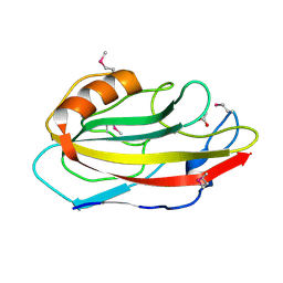
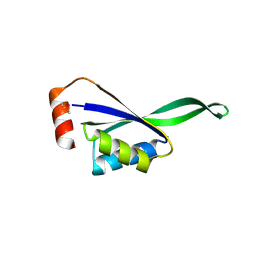
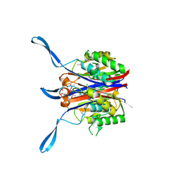
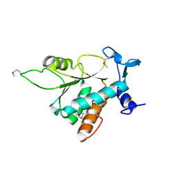
2N7D
DownloadVisualize
BU of 2n7d by Molmil
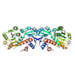
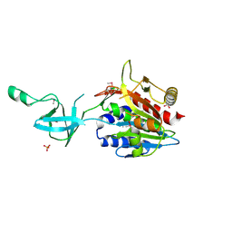
Solution structure of the UBL domain of human Ddi2
Descriptor: Protein DDI1 homolog 2
Authors:Siva, M, Grantz Saskova, K, Veverka, V.
Deposit date:2015-09-08
Release date:2016-07-20
Last modified:2024-05-15
Method:SOLUTION NMR
Cite:Human DNA-Damage-Inducible 2 Protein Is Structurally and Functionally Distinct from Its Yeast Ortholog.
Sci Rep, 6, 2016
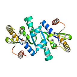
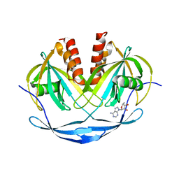
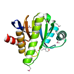
1WUT
DownloadVisualize
BU of 1wut by Molmil
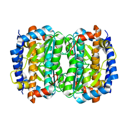
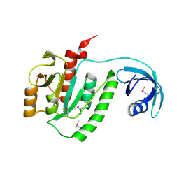
Acyl Ureas as Human Liver Glycogen Phosphorylase Inhibitors for the Treatment of Type 2 Diabetes
Descriptor: 7-[2,6-DICHLORO-4-({[(2-CHLOROBENZOYL)AMINO]CARBONYL}AMINO)PHENOXY]HEPTANOIC ACID, Glycogen phosphorylase, muscle form, ...
Authors:Klabunde, T, Wendt, K.U, Kadereit, D, Brachvogel, V, Burger, H.-J, Herling, A.W, Oikonomakos, N.G, Kosmopoulou, M.N, Schmoll, D, Sarubbi, E, von Roedern, E, Schonafinger, K, Defossa, E.
Deposit date:2004-12-08
Release date:2005-12-08
Last modified:2011-07-13
Method:X-RAY DIFFRACTION (2.26 Å)
Cite:Crystallographic studies on acyl ureas, a new class of glycogen phosphorylase inhibitors, as potential antidiabetic drugs
Protein Sci., 14, 2005
4LY1
DownloadVisualize
BU of 4ly1 by Molmil
Structure of Human HDAC2 in complex with inhibitor 4-(acetylamino)-N-[2-amino-5-(thiophen-2-yl)phenyl]benzamide
Descriptor: 2-[N-CYCLOHEXYLAMINO]ETHANE SULFONIC ACID, 4-(acetylamino)-N-[2-amino-5-(thiophen-2-yl)phenyl]benzamide, CALCIUM ION, ...
Authors:Fong, R, Lupardus, P.J.
Deposit date:2013-07-30
Release date:2013-08-21
Last modified:2023-09-20
Method:X-RAY DIFFRACTION (1.57 Å)
Cite:Histone Deacetylase (HDAC) Inhibitor Kinetic Rate Constants Correlate with Cellular Histone Acetylation but Not Transcription and Cell Viability.
J.Biol.Chem., 288, 2013
2VSY
DownloadVisualize
BU of 2vsy by Molmil
Xanthomonas campestris putative OGT (XCC0866), apostructure
Descriptor: 2-[N-CYCLOHEXYLAMINO]ETHANE SULFONIC ACID, CHLORIDE ION, DI(HYDROXYETHYL)ETHER, ...
Authors:Schuettelkopf, A.W, Clarke, A.J, van Aalten, D.M.F.
Deposit date:2008-05-01
Release date:2008-11-18
Last modified:2024-05-08
Method:X-RAY DIFFRACTION (2.1 Å)
Cite:Structural Insights Into Mechanism and Specificity of O-Glcnac Transferase.
Embo J., 27, 2008
7ZXK
DownloadVisualize
BU of 7zxk by Molmil
Human IL-27 in complex with neutralizing SRF388 FAb fragment
Descriptor: 2-acetamido-2-deoxy-beta-D-glucopyranose, Interleukin-27 subunit alpha, Interleukin-27 subunit beta, ...
Authors:Bloch, Y, Skladanowska, K, Strand, J, Welin, M, Logan, D, Hill, J, Savvides, S.N.
Deposit date:2022-05-21
Release date:2022-11-02
Last modified:2024-01-31
Method:X-RAY DIFFRACTION (2.2 Å)
Cite:Structural basis of activation and antagonism of receptor signaling mediated by interleukin-27.
Cell Rep, 41, 2022
2XVP
DownloadVisualize
BU of 2xvp by Molmil
ChiA1 from Aspergillus fumigatus, apostructure
Descriptor: CLASS III CHITINASE CHIA1, PHOSPHATE ION
Authors:Schuettelkopf, A.W, van Aalten, D.M.F.
Deposit date:2010-10-27
Release date:2011-06-01
Last modified:2023-12-20
Method:X-RAY DIFFRACTION (2 Å)
Cite:Natural Product-Guided Discovery of a Fungal Chitinase Inhibitor.
Chem.Biol., 17, 2010
7ZG0
DownloadVisualize
BU of 7zg0 by Molmil
Murine IL-27 in complex with IL-27Ra and a non-competing Nb
Descriptor: 2-acetamido-2-deoxy-beta-D-glucopyranose, 2-acetamido-2-deoxy-beta-D-glucopyranose-(1-4)-2-acetamido-2-deoxy-beta-D-glucopyranose, Interleukin-27 receptor subunit alpha, ...
Authors:Skladanowska, K, Bloch, Y, Savvides, S.N.
Deposit date:2022-04-01
Release date:2022-11-02
Last modified:2024-05-01
Method:X-RAY DIFFRACTION (3.18 Å)
Cite:Structural basis of activation and antagonism of receptor signaling mediated by interleukin-27.
Cell Rep, 41, 2022
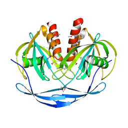
1VHN
DownloadVisualize
BU of 1vhn by Molmil
Crystal structure of a putative flavin oxidoreductase with flavin
Descriptor: FLAVIN MONONUCLEOTIDE, SULFATE ION, putative flavin oxidoreductase
Authors:Structural GenomiX
Deposit date:2003-12-01
Release date:2003-12-30
Last modified:2023-12-27
Method:X-RAY DIFFRACTION (1.59 Å)
Cite:The 1.59 A resolution crystal structure of TM0096, a flavin mononucleotide binding protein from Thermotoga maritima.
Proteins, 55, 2004
1VIO
DownloadVisualize
BU of 1vio by Molmil
Crystal structure of pseudouridylate synthase
Descriptor: 1,4-BUTANEDIOL, Ribosomal small subunit pseudouridine synthase A
Authors:Structural GenomiX
Deposit date:2003-12-01
Release date:2003-12-30
Last modified:2023-12-27
Method:X-RAY DIFFRACTION (1.59 Å)
Cite:Structure of the pseudouridine synthase RsuA from Haemophilus influenzae.
Acta Crystallogr.,Sect.F, 61, 2005
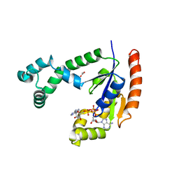
4OQX
DownloadVisualize
BU of 4oqx by Molmil
Crystal structure of thymidine kinase from herpes simplex virus type 1 in complex with Me-ARA-EdU
Descriptor: 1-(2-deoxy-2-methyl-beta-D-arabinofuranosyl)-5-ethynylpyrimidine-2,4(1H,3H)-dione, SULFATE ION, Thymidine kinase
Authors:Pernot, L, Neef, A.B, Westermaier, Y, Perozzo, R, Luedtke, N, Scapozza, L.
Deposit date:2014-02-10
Release date:2014-08-13
Last modified:2023-09-20
Method:X-RAY DIFFRACTION (2.5 Å)
Cite:Crystal structure of HSV1-TK complexed with Me-ARA-EdU
To be Published
4A5U
DownloadVisualize
BU of 4a5u by Molmil
Turnip yellow mosaic virus proteinase and Escherichia coli 30S ribosomal S15
Descriptor: 30S RIBOSOMAL PROTEIN S15, RNA REPLICASE POLYPROTEIN
Authors:Robin, C, Beaurepaire, L, Bressanelli, S.
Deposit date:2011-10-28
Release date:2012-11-14
Last modified:2024-05-08
Method:X-RAY DIFFRACTION (2 Å)
Cite:A Compact Viral Processing Proteinase/Ubiquitin Hydrolase from the Otu Family.
Plos Pathog., 9, 2013
2HX6
DownloadVisualize
BU of 2hx6 by Molmil
Solution structure analysis of the phage T4 endoribonuclease RegB
Descriptor: Ribonuclease
Authors:Odaert, B, Saida, F, Uzan, M, Bontems, F.
Deposit date:2006-08-02
Release date:2006-10-31
Last modified:2024-05-29
Method:SOLUTION NMR
Cite:Structural and functional studies of RegB, a new member of a family of sequence-specific ribonucleases involved in mRNA inactivation on the ribosome.
J.Biol.Chem., 282, 2007
3DK3
DownloadVisualize
BU of 3dk3 by Molmil
Crystal structure of mutant ABL kinase domain in complex with small molecule fragment
Descriptor: 2-amino-5-[3-(1-ethyl-1H-pyrazol-5-yl)-1H-pyrrolo[2,3-b]pyridin-5-yl]-N,N-dimethylbenzamide, Proto-oncogene tyrosine-protein kinase ABL1, SULFATE ION
Authors:Lewis, H.A.
Deposit date:2008-06-24
Release date:2008-07-29
Last modified:2024-02-21
Method:X-RAY DIFFRACTION (2.02 Å)
Cite:Crystal structure of mutant ABL kinase domain in complex with small molecule fragment
To be Published
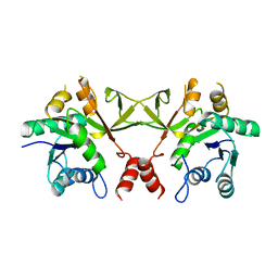
1O6B
DownloadVisualize
BU of 1o6b by Molmil
Crystal structure of phosphopantetheine adenylyltransferase with ADP
Descriptor: ADENOSINE-5'-DIPHOSPHATE, CHLORIDE ION, MAGNESIUM ION, ...
Authors:Structural GenomiX
Deposit date:2003-11-03
Release date:2003-11-25
Last modified:2023-12-27
Method:X-RAY DIFFRACTION (2.2 Å)
Cite:Structural analysis of a set of proteins resulting from a bacterial genomics project
Proteins, 60, 2005
1O63
DownloadVisualize
BU of 1o63 by Molmil
Crystal structure of an ATP phosphoribosyltransferase
Descriptor: ATP phosphoribosyltransferase
Authors:Structural GenomiX
Deposit date:2003-10-23
Release date:2003-11-11
Last modified:2023-12-27
Method:X-RAY DIFFRACTION (2 Å)
Cite:Structural analysis of a set of proteins resulting from a bacterial genomics project
Proteins, 60, 2005
1O6C
DownloadVisualize
BU of 1o6c by Molmil
Crystal structure of UDP-N-acetylglucosamine 2-epimerase
Descriptor: UDP-N-acetylglucosamine 2-epimerase
Authors:Structural GenomiX
Deposit date:2003-11-03
Release date:2003-11-25
Last modified:2023-12-27
Method:X-RAY DIFFRACTION (2.9 Å)
Cite:Structural analysis of a set of proteins resulting from a bacterial genomics project
Proteins, 60, 2005
1O60
DownloadVisualize
BU of 1o60 by Molmil
Crystal structure of KDO-8-phosphate synthase
Descriptor: 2-dehydro-3-deoxyphosphooctonate aldolase
Authors:Structural GenomiX
Deposit date:2003-10-23
Release date:2003-11-11
Last modified:2023-12-27
Method:X-RAY DIFFRACTION (1.8 Å)
Cite:Structural analysis of a set of proteins resulting from a bacterial genomics project
Proteins, 60, 2005
1O67
DownloadVisualize
BU of 1o67 by Molmil
Crystal structure of an hypothetical protein
Descriptor: Hypothetical protein yiiM
Authors:Structural GenomiX
Deposit date:2003-10-23
Release date:2003-11-11
Last modified:2023-12-27
Method:X-RAY DIFFRACTION (2.54 Å)
Cite:Structural analysis of a set of proteins resulting from a bacterial genomics project
Proteins, 60, 2005
1O61
DownloadVisualize
BU of 1o61 by Molmil
Crystal structure of a PLP-dependent enzyme with PLP
Descriptor: ACETATE ION, PYRIDOXAL-5'-PHOSPHATE, aminotransferase
Authors:Structural GenomiX
Deposit date:2003-10-23
Release date:2003-11-11
Last modified:2023-12-27
Method:X-RAY DIFFRACTION (1.9 Å)
Cite:Structural analysis of a set of proteins resulting from a bacterial genomics project
Proteins, 60, 2005
1O68
DownloadVisualize
BU of 1o68 by Molmil
Crystal structure of 3-methyl-2-oxobutanoate hydroxymethyltransferase
Descriptor: 3-METHYL-2-OXOBUTANOIC ACID, 3-methyl-2-oxobutanoate hydroxymethyltransferase, SODIUM ION
Authors:Structural GenomiX
Deposit date:2003-10-23
Release date:2003-11-11
Last modified:2023-12-27
Method:X-RAY DIFFRACTION (2.1 Å)
Cite:Structural analysis of a set of proteins resulting from a bacterial genomics project
Proteins, 60, 2005
3DK6
DownloadVisualize
BU of 3dk6 by Molmil
Crystal structure of mutant ABL kinase domain in complex with small molecule fragment
Descriptor: 2-amino-5-[3-(1-ethyl-1H-pyrazol-5-yl)-1H-pyrrolo[2,3-b]pyridin-5-yl]-N,N-dimethylbenzamide, Proto-oncogene tyrosine-protein kinase ABL1
Authors:Lewis, H.A.
Deposit date:2008-06-24
Release date:2008-07-29
Last modified:2021-10-20
Method:X-RAY DIFFRACTION (2.02 Å)
Cite:Crystal structure of mutant ABL kinase domain in complex with small molecule fragment
To be Published
1O6D
DownloadVisualize
BU of 1o6d by Molmil
Crystal structure of a hypothetical protein
Descriptor: Hypothetical UPF0247 protein TM0844
Authors:Structural GenomiX
Deposit date:2003-11-03
Release date:2003-11-25
Last modified:2023-12-27
Method:X-RAY DIFFRACTION (1.66 Å)
Cite:Structural analysis of a set of proteins resulting from a bacterial genomics project
Proteins, 60, 2005
1O7A
DownloadVisualize
BU of 1o7a by Molmil
Human beta-Hexosaminidase B
Descriptor: 1,2-ETHANEDIOL, 2-(acetylamido)-2-deoxy-D-glucono-1,5-lactone, 2-acetamido-2-deoxy-beta-D-glucopyranose, ...
Authors:Maier, T, Strater, N, Schuette, C, Klingenstein, R, Sandhoff, K, Saenger, W.
Deposit date:2002-10-29
Release date:2003-10-23
Last modified:2020-11-18
Method:X-RAY DIFFRACTION (2.25 Å)
Cite:The X-Ray Crystal Structure of Human Beta-Hexosaminidase B Provides New Insights Into Sandhoff Disease
J.Mol.Biol., 328, 2003

221716

건을2024-06-26부터공개중

PDB statisticsPDBj update infoContact PDBjnumon