Loading
PDBj
MenuPDBj@FacebookPDBj@TwitterPDBj@YouTubewwPDB FoundationwwPDB
RCSB PDBPDBeBMRBAdv. SearchSearch help
Search by PDB author
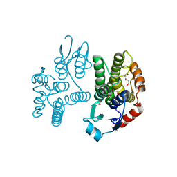
5AN1
DownloadVisualize
BU of 5an1 by Molmil
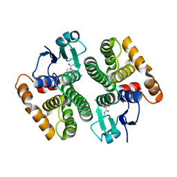
Crystallographic structure of the Glutathione S-Transferase from Litopenaeus vannamei complexed with Glutathione
Descriptor: GLUTATHIONE, GLUTATHIONE S-TRANSFERASE
Authors:Juarez-Martinez, A.B, Sotelo-Mundo, R, Rudino-Pinera, E.
Deposit date:2015-09-02
Release date:2016-10-12
Last modified:2024-01-10
Method:X-RAY DIFFRACTION (2 Å)
Cite:Crystal structure of a class-mu glutathione S-transferase from whiteleg shrimp Litopenaeus vannamei: structural changes in the xenobiotic binding H-site may alter the spectra of molecules bound.
J. Biochem. Mol. Toxicol., 31, 2017
5KEJ
DownloadVisualize
BU of 5kej by Molmil
Crystallographic structure of the Tau class glutathione S-transferase MiGSTU in complex with S-hexyl-glutathione
Descriptor: DI(HYDROXYETHYL)ETHER, S-HEXYLGLUTATHIONE, Tau class glutathione S-transferase
Authors:Valenzuela-Chavira, I, Serrano-Posada, H, Lopez-Zavala, A, Hernandez-Paredes, J, Sotelo-Mundo, R.
Deposit date:2016-06-09
Release date:2017-02-01
Last modified:2023-09-27
Method:X-RAY DIFFRACTION (2.35 Å)
Cite:Insights into ligand binding to a glutathione S-transferase from mango: Structure, thermodynamics and kinetics.
Biochimie, 135, 2017
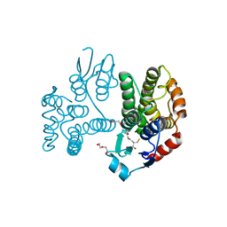
5G5E
DownloadVisualize
BU of 5g5e by Molmil
Crystallographic structure of the Tau class glutathione S-transferase MiGSTU from mango Mangifera indica L.
Descriptor: ACETATE ION, DI(HYDROXYETHYL)ETHER, TAU CLASS GLUTATHIONE S-TRANSFERASE
Authors:Valenzuela-Chavira, I, Serrano-Posada, H, Lopez-Zavala, A, Hernandez-Paredes, J, Sotelo-Mundo, R.
Deposit date:2016-05-24
Release date:2017-02-01
Last modified:2024-01-10
Method:X-RAY DIFFRACTION (1.8 Å)
Cite:Insights Into Ligand Binding to a Glutathione S-Transferase from Mango: Structure, Thermodynamics and Kinetics
Biochimie, 135, 2017
5G5F
DownloadVisualize
BU of 5g5f by Molmil
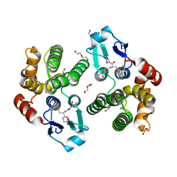
Crystallographic structure of the Tau class glutathione S-transferase MiGSTU in complex with reduced glutathione.
Descriptor: DI(HYDROXYETHYL)ETHER, GLUTATHIONE, TAU CLASS GLUTATHIONE S-TRANSFERASE
Authors:Valenzuela-Chavira, I, Serrano-Posada, H, Lopez-Zavala, A, Hernandez-Paredes, J, Sotelo-Mundo, R.
Deposit date:2016-05-24
Release date:2017-02-01
Last modified:2024-01-10
Method:X-RAY DIFFRACTION (2.3 Å)
Cite:Insights Into Ligand Binding to a Glutathione S-Transferase from Mango: Structure, Thermodynamics and Kinetics
Biochimie, 135, 2017
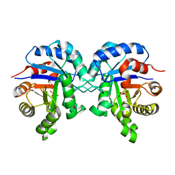
6BVE
DownloadVisualize
BU of 6bve by Molmil
Triosephosphate isomerase of Synechocystis in complex with 2-Phosphoglycolic acid
Descriptor: 2-PHOSPHOGLYCOLIC ACID, SODIUM ION, Triosephosphate isomerase
Authors:Jimenez-Sandoval, P, Castro-Torres, E, Brieba, L.G.
Deposit date:2017-12-12
Release date:2018-12-19
Last modified:2023-10-04
Method:X-RAY DIFFRACTION (1.78 Å)
Cite:Structural Basis for the Limited Response to Oxidative and Thiol-Conjugating Agents by Triosephosphate Isomerase From the Photosynthetic BacteriaSynechocystis.
Front Mol Biosci, 5, 2018
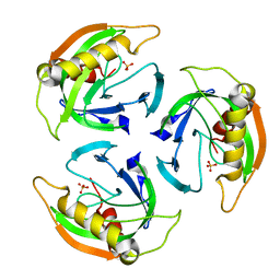
4UM4
DownloadVisualize
BU of 4um4 by Molmil
STRUCTURE OF INORGANIC PYROPHOSPHATASE FROM ESCHERICHIA COLI IN COMPLEX WITH SULFATE
Descriptor: INORGANIC PYROPHOSPHATASE, SULFATE ION
Authors:Campos-Acevedo, A.A, Rudino-Pinera, E.
Deposit date:2014-05-15
Release date:2014-11-26
Last modified:2024-01-10
Method:X-RAY DIFFRACTION (2.65 Å)
Cite:First attempts to crystallize a non-homogeneous sample of thioredoxin from Litopenaeus vannamei: What to do when you have diffraction data of a protein that is not the target?
Biochem Biophys Rep, 8, 2016
6UL3
DownloadVisualize
BU of 6ul3 by Molmil
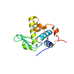
Crystal structure of a lysozyme from Litopenaeus vannamei
Descriptor: Lysozyme
Authors:Hernandez-Santoyo, A, Rodriguez-Romero, A.
Deposit date:2019-10-06
Release date:2020-03-25
Last modified:2024-10-16
Method:X-RAY DIFFRACTION (3 Å)
Cite:Crystal structure of a C-type lysozyme from Litopenaeus vanamei exhibiting a high binding constant to its chitotriose inhibitor.
Fish Shellfish Immunol., 100, 2020
6UKC
DownloadVisualize
BU of 6ukc by Molmil
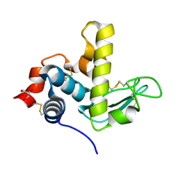
Crystal structure of a lysozyme from Litopenaeus vannamei
Descriptor: GLYCEROL, Lysozyme
Authors:Hernandez-Santoyo, A, Rodriguez-Romero, A.
Deposit date:2019-10-04
Release date:2020-03-25
Last modified:2024-11-06
Method:X-RAY DIFFRACTION (2.25 Å)
Cite:Crystal structure of a C-type lysozyme from Litopenaeus vanamei exhibiting a high binding constant to its chitotriose inhibitor.
Fish Shellfish Immunol., 100, 2020
5G2Z
DownloadVisualize
BU of 5g2z by Molmil
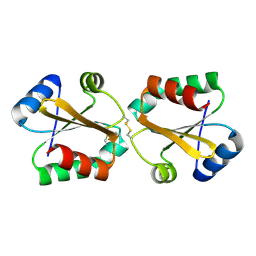
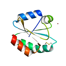
Crystallographic structure of mutant W31A of thioredoxin from Litopenaeus vannamei
Descriptor: THIOREDOXIN
Authors:Campos-Acevedo, A.A, Rudino-Pinera, E.
Deposit date:2016-04-18
Release date:2017-03-15
Last modified:2024-10-16
Method:X-RAY DIFFRACTION (1.88 Å)
Cite:Is dimerization a common feature in thioredoxins? The case of thioredoxin from Litopenaeus vannamei.
Acta Crystallogr D Struct Biol, 73, 2017
5G30
DownloadVisualize
BU of 5g30 by Molmil
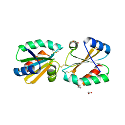
Crystallographic structure of mutant D60S of thioredoxin from Litopenaeus vannamei
Descriptor: ACETATE ION, DI(HYDROXYETHYL)ETHER, THIOREDOXIN
Authors:Campos-Acevedo, A.A, Rudino-Pinera, E.
Deposit date:2016-04-18
Release date:2017-03-15
Last modified:2024-10-23
Method:X-RAY DIFFRACTION (1.65 Å)
Cite:Is dimerization a common feature in thioredoxins? The case of thioredoxin from Litopenaeus vannamei.
Acta Crystallogr D Struct Biol, 73, 2017
5G31
DownloadVisualize
BU of 5g31 by Molmil
Crystallographic structure of mutant C73S of thioredoxin from Litopenaeus vannamei
Descriptor: THIOREDOXIN, ZINC ION
Authors:Campos-Acevedo, A.A, Rudino-Pinera, E.
Deposit date:2016-04-18
Release date:2017-03-15
Last modified:2024-01-10
Method:X-RAY DIFFRACTION (2 Å)
Cite:Is dimerization a common feature in thioredoxins? The case of thioredoxin from Litopenaeus vannamei.
Acta Crystallogr D Struct Biol, 73, 2017
<12


227111

건을2024-11-06부터공개중

PDB statisticsPDBj update infoContact PDBjnumon