Loading
PDBj
MenuPDBj@FacebookPDBj@TwitterPDBj@YouTubewwPDB FoundationwwPDB
RCSB PDBPDBeBMRBAdv. SearchSearch help
Search by PDB author
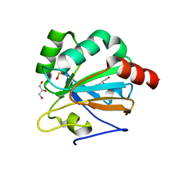
5ID2
DownloadVisualize
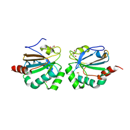
BU of 5id2 by Molmil
Asymmetry in the active site of Mycobacterium tuberculosis AhpE upon exposure to Mycothiol
Descriptor: ACETATE ION, GLYCEROL, Putative peroxiredoxin Rv2238c
Authors:Kumar, A, Balakrishna, A.M, Gruber, G.
Deposit date:2016-02-23
Release date:2016-08-03
Last modified:2024-10-23
Method:X-RAY DIFFRACTION (2.43 Å)
Cite:Redox chemistry of Mycobacterium tuberculosis alkylhydroperoxide reductase E (AhpE): Structural and mechanistic insight into a mycoredoxin-1 independent reductive pathway of AhpE via mycothiol
Free Radic. Biol. Med., 97, 2016
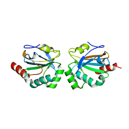
1B68
DownloadVisualize
BU of 1b68 by Molmil
APOLIPOPROTEIN E4 (APOE4), 22K FRAGMENT
Descriptor: APOLIPOPROTEIN E
Authors:Rupp, B, Peters-Libeu, C.
Deposit date:1999-01-21
Release date:2001-07-11
Last modified:2023-08-09
Method:X-RAY DIFFRACTION (2 Å)
Cite:Interaction of the N-terminal domain of apolipoprotein E4 with heparin.
Biochemistry, 40, 2001
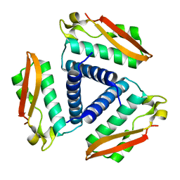
4X1U
DownloadVisualize
BU of 4x1u by Molmil
The structure of AhpE from Mycobacterium tuberculosis revisited
Descriptor: GLYCEROL, Putative peroxiredoxin MT2298
Authors:Pallo, A, Messens, J.
Deposit date:2014-11-25
Release date:2016-07-27
Last modified:2024-10-23
Method:X-RAY DIFFRACTION (1.87 Å)
Cite:Revisiting sulfur H-bonds in proteins: The example of peroxiredoxin AhpE.
Sci Rep, 6, 2016
4X0X
DownloadVisualize
BU of 4x0x by Molmil
The structure of AhpE from Mycobacterium tuberculosis revisited
Descriptor: Putative peroxiredoxin MT2298
Authors:Pallo, A, Messens, J.
Deposit date:2014-11-24
Release date:2016-07-27
Last modified:2024-01-10
Method:X-RAY DIFFRACTION (1.9 Å)
Cite:Revisiting sulfur H-bonds in proteins: The example of peroxiredoxin AhpE.
Sci Rep, 6, 2016
2FR2
DownloadVisualize
BU of 2fr2 by Molmil
Crystal Structure of Rv2717c from M. tuberculosis
Descriptor: hypothetical protein Rv2717c
Authors:Hung, L.W, TB Structural Genomics Consortium (TBSGC)
Deposit date:2006-01-18
Release date:2006-01-31
Last modified:2011-07-13
Method:X-RAY DIFFRACTION (1.5 Å)
Cite:Crystal structure of Rv2717c from Mycobacterium tuberculosis
To be Published
2FVH
DownloadVisualize
BU of 2fvh by Molmil
Crystal Structure of Rv1848, a Urease Gamma Subunit UreA (Urea amidohydrolase), from Mycobacterium Tuberculosis
Descriptor: Urease gamma subunit
Authors:Bursey, E.H, Yu, M, TB Structural Genomics Consortium (TBSGC), Lekin, T, Rupp, B, Kim, C.Y, Rho, B.S, Terwilliger, T.C.
Deposit date:2006-01-30
Release date:2006-02-07
Last modified:2024-02-14
Method:X-RAY DIFFRACTION (1.8 Å)
Cite:Crystal Structure of Rv1848, a Urease Gamma Subunit UreA (Urea amidohydrolase), from Mycobacterium Tuberculosis
To be Published
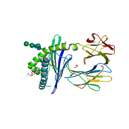
1ZHN
DownloadVisualize
BU of 1zhn by Molmil
Crystal Structure of mouse CD1d bound to the self ligand phosphatidylcholine
Descriptor: 2-acetamido-2-deoxy-beta-D-glucopyranose, 7-[(DODECANOYLOXY)METHYL]-4-HYDROXY-N,N,N-TRIMETHYL-9-OXO-3,5,8-TRIOXA-4-PHOSPHADOTRIACONTAN-1-AMINIUM 4-OXIDE, CD1d1 antigen, ...
Authors:Giabbai, B, Sidobre, S, Crispin, M.M.D, Sanchez Ruiz, Y, Bachi, A, Kronenberg, M, Wilson, I.A, Degano, M.
Deposit date:2005-04-26
Release date:2005-07-19
Last modified:2024-10-30
Method:X-RAY DIFFRACTION (2.8 Å)
Cite:Crystal structure of mouse CD1d bound to the self ligand phosphatidylcholine: a molecular basis for NKT cell activation
J.Immunol., 175, 2005
<12


227111

건을2024-11-06부터공개중

PDB statisticsPDBj update infoContact PDBjnumon