Loading
PDBj
MenuPDBj@FacebookPDBj@TwitterPDBj@YouTubewwPDB FoundationwwPDB
RCSB PDBPDBeBMRBAdv. SearchSearch help
Search by PDB author
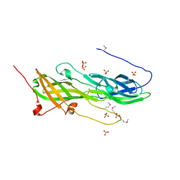
3NPD
DownloadVisualize
BU of 3npd by Molmil
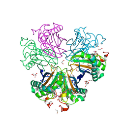
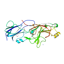
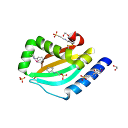
Crystal structure of a putative secreted protein (PA3611) from PSEUDOMONAS AERUGINOSA at 1.60 A resolution
Descriptor: 1,2-ETHANEDIOL, 3-CYCLOHEXYL-1-PROPYLSULFONIC ACID, SULFATE ION, ...
Authors:Joint Center for Structural Genomics (JCSG)
Deposit date:2010-06-28
Release date:2010-07-28
Last modified:2023-02-01
Method:X-RAY DIFFRACTION (1.6 Å)
Cite:Crystal structure of a putative quorum sensing-regulated protein (PA3611) from the Pseudomonas-specific DUF4146 family.
Proteins, 82, 2014
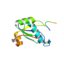
3NL9
DownloadVisualize
BU of 3nl9 by Molmil
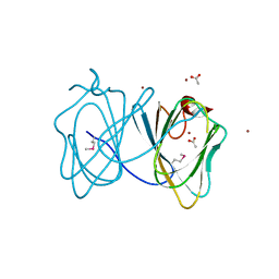
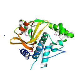
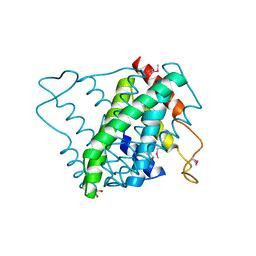
Crystal structure of a putative NTP pyrophosphohydrolase (Exig_1061) from EXIGUOBACTERIUM SP. 255-15 at 1.78 A resolution
Descriptor: 1,2-ETHANEDIOL, putative NTP pyrophosphohydrolase
Authors:Joint Center for Structural Genomics (JCSG)
Deposit date:2010-06-21
Release date:2010-07-21
Last modified:2023-02-01
Method:X-RAY DIFFRACTION (1.78 Å)
Cite:Structure of a putative NTP pyrophosphohydrolase: YP_001813558.1 from Exiguobacterium sibiricum 255-15.
Acta Crystallogr.,Sect.F, 66, 2010
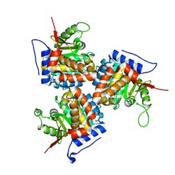
3BYQ
DownloadVisualize
BU of 3byq by Molmil
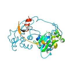
Crystal structure of a duf1185 family protein (bb2672) from bordetella bronchiseptica rb50 at 1.70 A resolution
Descriptor: 1,2-ETHANEDIOL, CHLORIDE ION, TETRAETHYLENE GLYCOL, ...
Authors:Joint Center for Structural Genomics (JCSG)
Deposit date:2008-01-16
Release date:2008-01-29
Last modified:2023-02-01
Method:X-RAY DIFFRACTION (1.7 Å)
Cite:Structures of the first representatives of Pfam family PF06684 (DUF1185) reveal a novel variant of the Bacillus chorismate mutase fold and suggest a role in amino-acid metabolism.
Acta Crystallogr.,Sect.F, 66, 2010
3H50
DownloadVisualize
BU of 3h50 by Molmil
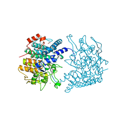
CRYSTAL STRUCTURE OF A TETRACENOMYCIN POLYKETIDE SYNTHESIS PROTEIN (TCMJ) FROM XANTHOMONAS CAMPESTRIS PV. CAMPESTRIS AT 1.60 A RESOLUTION
Descriptor: ACETATE ION, Tetracenomycin polyketide synthesis protein, ZINC ION
Authors:Joint Center for Structural Genomics (JCSG)
Deposit date:2009-04-21
Release date:2009-05-05
Last modified:2023-02-01
Method:X-RAY DIFFRACTION (1.6 Å)
Cite:Conformational changes associated with the binding of zinc acetate at the putative active site of XcTcmJ, a cupin from Xanthomonas campestris pv. campestris.
Acta Crystallogr.,Sect.F, 66, 2010
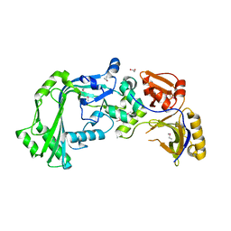
3D00
DownloadVisualize
BU of 3d00 by Molmil
Crystal structure of a tungsten formylmethanofuran dehydrogenase subunit e (fmde)-like protein (syn_00638) from syntrophus aciditrophicus at 1.90 A resolution
Descriptor: CHLORIDE ION, Tungsten formylmethanofuran dehydrogenase subunit E, ZINC ION
Authors:Joint Center for Structural Genomics (JCSG)
Deposit date:2008-04-30
Release date:2008-05-20
Last modified:2023-02-01
Method:X-RAY DIFFRACTION (1.9 Å)
Cite:Structures of three members of Pfam PF02663 (FmdE) implicated in microbial methanogenesis reveal a conserved alpha+beta core domain and an auxiliary C-terminal treble-clef zinc finger.
Acta Crystallogr.,Sect.F, 66, 2010
3CGH
DownloadVisualize
BU of 3cgh by Molmil
Crystal structure of a susd homolog (bt_3984) from bacteroides thetaiotaomicron vpi-5482 at 1.70 A resolution
Descriptor: ACETATE ION, SusD homolog, ZINC ION
Authors:Joint Center for Structural Genomics (JCSG)
Deposit date:2008-03-05
Release date:2008-03-18
Last modified:2023-02-01
Method:X-RAY DIFFRACTION (1.7 Å)
Cite:Structure of BT_3984, a member of the SusD/RagB family of nutrient-binding molecules.
Acta Crystallogr.,Sect.F, 66, 2010
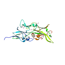
5CAG
DownloadVisualize
BU of 5cag by Molmil
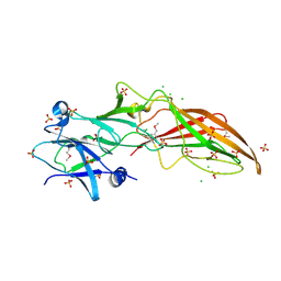
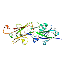
Crystal structure of a putative adhesin (BACOVA_02677) from Bacteroides ovatus ATCC 8483 at 3.00 A resolution (PSI Community Target, Nakayama)
Descriptor: SULFATE ION, Uncharacterized protein
Authors:Joint Center for Structural Genomics (JCSG), Nakayama, K.
Deposit date:2015-06-29
Release date:2015-10-14
Last modified:2020-04-22
Method:X-RAY DIFFRACTION (3 Å)
Cite:A Distinct Type of Pilus from the Human Microbiome.
Cell, 165, 2016
5CXT
DownloadVisualize
BU of 5cxt by Molmil
Crystal structure of a RNA-binding protein 39 (RBM39) in complex with fragment of splicing factor (U2AF) from Unknown at 2.20 A resolution
Descriptor: RNA-binding protein 39, Splicing factor U2AF 65 kDa subunit
Authors:Joint Center for Structural Genomics (JCSG), Partnership for T-Cell Biology (TCELL)
Deposit date:2015-07-29
Release date:2015-10-14
Last modified:2023-09-27
Method:X-RAY DIFFRACTION (2.2 Å)
Cite:UHM-ULM interactions in the RBM39-U2AF65 splicing-factor complex.
Acta Crystallogr D Struct Biol, 72, 2016
1ESJ
DownloadVisualize
BU of 1esj by Molmil
CRYSTAL STRUCTURE OF THIAZOLE KINASE MUTANT (C198S)
Descriptor: HYDROXYETHYLTHIAZOLE KINASE, SULFATE ION
Authors:Campobasso, N, Mathews, I.I, Begley, T.P, Ealick, S.E.
Deposit date:2000-04-10
Release date:2000-08-09
Last modified:2024-02-07
Method:X-RAY DIFFRACTION (1.8 Å)
Cite:Crystal structure of 4-methyl-5-beta-hydroxyethylthiazole kinase from Bacillus subtilis at 1.5 A resolution.
Biochemistry, 39, 2000
3KK7
DownloadVisualize
BU of 3kk7 by Molmil
Crystal structure of Putative cell invasion protein with MAC/Perforin domain (NP_812351.1) from BACTERIODES THETAIOTAOMICRON VPI-5482 at 2.46 A resolution
Descriptor: (4S)-2-METHYL-2,4-PENTANEDIOL, 1,2-ETHANEDIOL, CHLORIDE ION, ...
Authors:Joint Center for Structural Genomics (JCSG)
Deposit date:2009-11-04
Release date:2009-11-24
Last modified:2023-02-01
Method:X-RAY DIFFRACTION (2.46 Å)
Cite:Structure of a membrane-attack complex/perforin (MACPF) family protein from the human gut symbiont Bacteroides thetaiotaomicron.
Acta Crystallogr.,Sect.F, 66, 2010
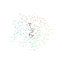
1G69
DownloadVisualize
BU of 1g69 by Molmil
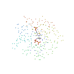
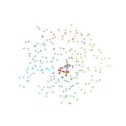
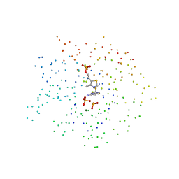
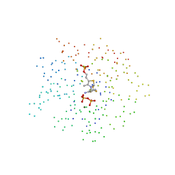
THIAMIN PHOSPHATE SYNTHASE
Descriptor: 2-METHYL-5-METHYLENE-5H-PYRIMIDIN-4-YLIDENEAMINE, 4-METHYL-5-HYDROXYETHYLTHIAZOLE PHOSPHATE, MAGNESIUM ION, ...
Authors:Peapus, D.H, Chiu, H.-J, Campobasso, N, Reddick, J.J, Begley, T.P, Ealick, S.E.
Deposit date:2000-11-03
Release date:2001-09-26
Last modified:2023-08-09
Method:X-RAY DIFFRACTION (1.5 Å)
Cite:Structural characterization of the enzyme-substrate, enzyme-intermediate, and enzyme-product complexes of thiamin phosphate synthase.
Biochemistry, 40, 2001
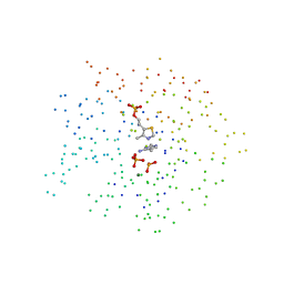
1G4P
DownloadVisualize
BU of 1g4p by Molmil
THIAMIN PHOSPHATE SYNTHASE
Descriptor: 4-AMINO-2-TRIFLUOROMETHYL-5-HYDROXYMETHYLPYRIMIDINE PYROPHOSPHATE, MAGNESIUM ION, THIAMIN PHOSPHATE SYNTHASE
Authors:Peapus, D.H, Chiu, H.-J, Campobasso, N, Reddick, J.J, Begley, T.P, Ealick, S.E.
Deposit date:2000-10-27
Release date:2001-09-26
Last modified:2023-08-09
Method:X-RAY DIFFRACTION (2.5 Å)
Cite:Structural characterization of the enzyme-substrate, enzyme-intermediate, and enzyme-product complexes of thiamin phosphate synthase.
Biochemistry, 40, 2001
1G6C
DownloadVisualize
BU of 1g6c by Molmil
THIAMIN PHOSPHATE SYNTHASE
Descriptor: 2-TRIFLUOROMETHYL-5-METHYLENE-5H-PYRIMIDIN-4-YLIDENEAMINE, 4-METHYL-5-HYDROXYETHYLTHIAZOLE PHOSPHATE, MAGNESIUM ION, ...
Authors:Peapus, D.H, Chiu, H.-J, Campobasso, N, Reddick, J.J, Begley, T.P, Ealick, S.E.
Deposit date:2000-11-03
Release date:2001-09-26
Last modified:2023-08-09
Method:X-RAY DIFFRACTION (1.4 Å)
Cite:Structural characterization of the enzyme-substrate, enzyme-intermediate, and enzyme-product complexes of thiamin phosphate synthase.
Biochemistry, 40, 2001
1G4E
DownloadVisualize
BU of 1g4e by Molmil
THIAMIN PHOSPHATE SYNTHASE
Descriptor: THIAMIN PHOSPHATE SYNTHASE
Authors:Peapus, D.H, Chiu, H.-J, Campobasso, N, Reddick, J.J, Begley, T.P, Ealick, S.E.
Deposit date:2000-10-26
Release date:2001-09-26
Last modified:2023-08-09
Method:X-RAY DIFFRACTION (1.6 Å)
Cite:Structural characterization of the enzyme-substrate, enzyme-intermediate, and enzyme-product complexes of thiamin phosphate synthase.
Biochemistry, 40, 2001
1G4T
DownloadVisualize
BU of 1g4t by Molmil
THIAMIN PHOSPHATE SYNTHASE
Descriptor: 3-(4-AMINO-2-TRIFLUOROMETHYL-PYRIMIDIN-5-YLMETHYL)-4-METHYL-5-(2-PHOSPHONATOOXY-ETHYL)-THIAZOL-3-IUM, MAGNESIUM ION, PYROPHOSPHATE 2-, ...
Authors:Peapus, D.H, Chiu, H.-J, Campobasso, N, Reddick, J.J, Begley, T.P, Ealick, S.E.
Deposit date:2000-10-28
Release date:2001-09-26
Last modified:2023-08-09
Method:X-RAY DIFFRACTION (1.55 Å)
Cite:Structural characterization of the enzyme-substrate, enzyme-intermediate, and enzyme-product complexes of thiamin phosphate synthase.
Biochemistry, 40, 2001
1G4S
DownloadVisualize
BU of 1g4s by Molmil
THIAMIN PHOSPHATE SYNTHASE
Descriptor: MAGNESIUM ION, PYROPHOSPHATE 2-, THIAMIN PHOSPHATE, ...
Authors:Peapus, D.H, Chiu, H.-J, Campobasso, N, Reddick, J.J, Begley, T.P, Ealick, S.E.
Deposit date:2000-10-27
Release date:2001-09-26
Last modified:2023-08-09
Method:X-RAY DIFFRACTION (1.7 Å)
Cite:Structural characterization of the enzyme-substrate, enzyme-intermediate, and enzyme-product complexes of thiamin phosphate synthase.
Biochemistry, 40, 2001
1G67
DownloadVisualize
BU of 1g67 by Molmil
THIAMIN PHOSPHATE SYNTHASE
Descriptor: 2-METHYL-5-METHYLENE-5H-PYRIMIDIN-4-YLIDENEAMINE, 4-METHYL-5-HYDROXYETHYLTHIAZOLE PHOSPHATE, MAGNESIUM ION, ...
Authors:Peapus, D.H, Chiu, H.-J, Campobasso, N, Reddick, J.J, Begley, T.P, Ealick, S.E.
Deposit date:2000-11-03
Release date:2001-09-26
Last modified:2023-08-09
Method:X-RAY DIFFRACTION (1.4 Å)
Cite:Structural characterization of the enzyme-substrate, enzyme-intermediate, and enzyme-product complexes of thiamin phosphate synthase.
Biochemistry, 40, 2001
4QDG
DownloadVisualize
BU of 4qdg by Molmil
Crystal structure of a putative adhesin (BT2657) from Bacteroides thetaiotaomicron VPI-5482 at 2.20 A resolution (PSI Community Target, Nakayama)
Descriptor: 1,2-ETHANEDIOL, MAGNESIUM ION, Putative adhesin
Authors:Joint Center for Structural Genomics (JCSG)
Deposit date:2014-05-13
Release date:2014-06-11
Last modified:2023-02-01
Method:X-RAY DIFFRACTION (2.2 Å)
Cite:A Distinct Type of Pilus from the Human Microbiome.
Cell, 165, 2016
4Q5K
DownloadVisualize
BU of 4q5k by Molmil
Crystal structure of a N-acetylmuramoyl-L-alanine amidase (BACUNI_02947) from Bacteroides uniformis ATCC 8492 at 1.30 A resolution
Descriptor: (2R)-2-[[(1R,2S,3R,4R,5R)-4-acetamido-2-[(2S,3R,4R,5S,6R)-3-acetamido-6-(hydroxymethyl)-4,5-bis(oxidanyl)oxan-2-yl]oxy-6,8-dioxabicyclo[3.2.1]octan-3-yl]oxy]propanoic acid, SODIUM ION, Uncharacterized protein
Authors:Joint Center for Structural Genomics (JCSG)
Deposit date:2014-04-17
Release date:2014-05-21
Last modified:2023-12-06
Method:X-RAY DIFFRACTION (1.3 Å)
Cite:Structure-guided functional characterization of DUF1460 reveals a highly specific NlpC/P60 amidase family.
Structure, 22, 2014
4Q98
DownloadVisualize
BU of 4q98 by Molmil
Crystal structure of a fimbrilin (fimA) from Porphyromonas gingivalis W83 at 1.30 A resolution (PSI Community Target, Nakayama)
Descriptor: Major fimbrial subunit protein, SODIUM ION
Authors:Joint Center for Structural Genomics (JCSG)
Deposit date:2014-04-29
Release date:2014-06-04
Last modified:2023-02-01
Method:X-RAY DIFFRACTION (1.3 Å)
Cite:A Distinct Type of Pilus from the Human Microbiome.
Cell, 165, 2016
4QB7
DownloadVisualize
BU of 4qb7 by Molmil
Crystal structure of a fimbrial protein (BVU_2522) from Bacteroides vulgatus ATCC 8482 at 2.55 A resolution
Descriptor: CHLORIDE ION, SULFATE ION, Uncharacterized protein
Authors:Joint Center for Structural Genomics (JCSG)
Deposit date:2014-05-06
Release date:2014-07-02
Last modified:2023-02-01
Method:X-RAY DIFFRACTION (2.55 Å)
Cite:A Distinct Type of Pilus from the Human Microbiome.
Cell, 165, 2016
4Q68
DownloadVisualize
BU of 4q68 by Molmil
Crystal structure of a N-acetylmuramoyl-L-alanine amidase (BACUNI_02947) from Bacteroides uniformis ATCC 8492 at 1.07 A resolution
Descriptor: 2-acetamido-2-deoxy-beta-D-glucopyranose, SODIUM ION, Uncharacterized protein
Authors:Joint Center for Structural Genomics (JCSG)
Deposit date:2014-04-21
Release date:2014-05-07
Last modified:2023-12-06
Method:X-RAY DIFFRACTION (1.07 Å)
Cite:Structure-guided functional characterization of DUF1460 reveals a highly specific NlpC/P60 amidase family.
Structure, 22, 2014
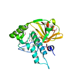
4QK4
DownloadVisualize
BU of 4qk4 by Molmil
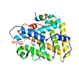
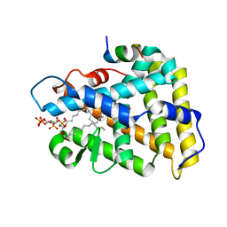
Crystal structure of human nuclear receptor sf-1 (nr5a1) bound to pip2 at 2.8 a resolution
Descriptor: (2S)-3-{[(R)-hydroxy{[(1R,2R,3S,4R,5R,6S)-2,3,6-trihydroxy-4,5-bis(phosphonooxy)cyclohexyl]oxy}phosphoryl]oxy}propane-1,2-diyl dihexadecanoate, 1,2-ETHANEDIOL, Peroxisome proliferator-activated receptor gamma coactivator 1-alpha, ...
Authors:Joint Center for Structural Genomics (JCSG), Partnership for Stem Cell Biology (STEMCELL)
Deposit date:2014-06-05
Release date:2014-07-30
Last modified:2024-03-13
Method:X-RAY DIFFRACTION (2.81 Å)
Cite:The signaling phospholipid PIP3 creates a new interaction surface on the nuclear receptor SF-1.
Proc.Natl.Acad.Sci.USA, 111, 2014
4QJR
DownloadVisualize
BU of 4qjr by Molmil
Crystal structure of human nuclear receptor sf-1 (nr5a1) bound to its hormone pip3 at 2.4 a resolution
Descriptor: (2S)-3-{[(R)-{[(1S,2S,3R,4S,5S,6S)-2,6-dihydroxy-3,4,5-tris(phosphonooxy)cyclohexyl]oxy}(hydroxy)phosphoryl]oxy}propane -1,2-diyl dihexadecanoate, ACETATE ION, Peroxisome proliferator-activated receptor gamma coactivator 1-alpha, ...
Authors:Joint Center for Structural Genomics (JCSG), Partnership for Stem Cell Biology (STEMCELL)
Deposit date:2014-06-04
Release date:2014-07-30
Last modified:2024-03-13
Method:X-RAY DIFFRACTION (2.4 Å)
Cite:The signaling phospholipid PIP3 creates a new interaction surface on the nuclear receptor SF-1.
Proc.Natl.Acad.Sci.USA, 111, 2014
4RDB
DownloadVisualize
BU of 4rdb by Molmil
Crystal structure of an immunoreactive 32 kDa antigen PG49 (PG_0181) from Porphyromonas gingivalis W83 at 1.45 A resolution (PSI Community Target, Nakayama)
Descriptor: 1,2-ETHANEDIOL, ACETONE, Immunoreactive 32 kDa antigen PG49
Authors:Joint Center for Structural Genomics (JCSG)
Deposit date:2014-09-18
Release date:2014-12-03
Last modified:2023-02-01
Method:X-RAY DIFFRACTION (1.45 Å)
Cite:A Distinct Type of Pilus from the Human Microbiome.
Cell, 165, 2016

222624

건을2024-07-17부터공개중

PDB statisticsPDBj update infoContact PDBjnumon