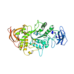
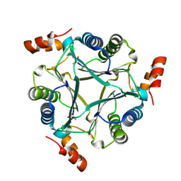
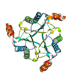
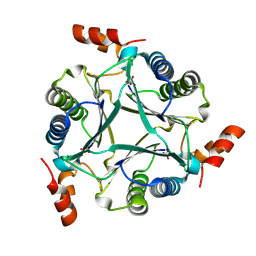
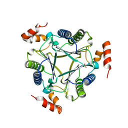
4RLF
| Crystal structure of a benzoate coenzyme A ligase with p-Toluic acid and o-Toluic acid | Descriptor: | 2-methylbenzoic acid, 4-METHYLBENZOIC ACID, Benzoate-coenzyme A ligase, ... | Authors: | Strom, S, Nosrati, M, Thornburg, C, Walker, K, Geiger, J.H. | Deposit date: | 2014-10-16 | Release date: | 2015-09-30 | Last modified: | 2023-09-20 | Method: | X-RAY DIFFRACTION (1.73 Å) | Cite: | Kinetically and Crystallographically Guided Mutations of a Benzoate CoA Ligase (BadA) Elucidate Mechanism and Expand Substrate Permissivity. Biochemistry, 54, 2015
|
|
4RM3
| Crystal structure of a benzoate coenzyme A ligase with 2-Furoic acid | Descriptor: | 2-FUROIC ACID, Benzoate-coenzyme A ligase | Authors: | Strom, S, Nosrati, M, Thornburg, C, Walker, K.D, Geiger, J.H. | Deposit date: | 2014-10-18 | Release date: | 2015-09-30 | Last modified: | 2023-09-20 | Method: | X-RAY DIFFRACTION (1.76 Å) | Cite: | Kinetically and Crystallographically Guided Mutations of a Benzoate CoA Ligase (BadA) Elucidate Mechanism and Expand Substrate Permissivity. Biochemistry, 54, 2015
|
|
4RUU
| |
4RMN
| Crystal structure of a benzoate coenzyme A ligase with 2-Thiophene Carboxylic acid | Descriptor: | Benzoate-coenzyme A ligase, GLYCEROL, THIOPHENE-2-CARBOXYLIC ACID | Authors: | Strom, S, Nosrati, M, Thornburg, C, Walker, K.D, Geiger, J.H. | Deposit date: | 2014-10-21 | Release date: | 2015-09-30 | Last modified: | 2023-09-20 | Method: | X-RAY DIFFRACTION (1.72 Å) | Cite: | Kinetically and Crystallographically Guided Mutations of a Benzoate CoA Ligase (BadA) Elucidate Mechanism and Expand Substrate Permissivity. Biochemistry, 54, 2015
|
|
4RM2
| Crystal structure of a benzoate coenzyme A ligase with 2-Fluoro benzoic acid | Descriptor: | 2-fluorobenzoic acid, Benzoate-coenzyme A ligase | Authors: | Strom, S, Nosrati, M, Thornburg, C, Walker, K.D, Geiger, J.H. | Deposit date: | 2014-10-18 | Release date: | 2015-09-30 | Last modified: | 2023-09-20 | Method: | X-RAY DIFFRACTION (1.77 Å) | Cite: | Kinetically and Crystallographically Guided Mutations of a Benzoate CoA Ligase (BadA) Elucidate Mechanism and Expand Substrate Permissivity. Biochemistry, 54, 2015
|
|
7ML5
| Structure of the Starch Branching Enzyme I (BEI) complexed with maltododecaose from Oryza sativa L | Descriptor: | Isoform 2 of 1,4-alpha-glucan-branching enzyme, chloroplastic/amyloplastic, alpha-D-glucopyranose-(1-4)-alpha-D-glucopyranose-(1-4)-alpha-D-glucopyranose-(1-4)-alpha-D-glucopyranose, ... | Authors: | Nayebi Gavgani, H, Fawaz, R, Geiger, J.H. | Deposit date: | 2021-04-27 | Release date: | 2021-11-17 | Last modified: | 2023-10-18 | Method: | X-RAY DIFFRACTION (2.35 Å) | Cite: | A structural explanation for the mechanism and specificity of plant branching enzymes I and IIb. J.Biol.Chem., 298, 2021
|
|
7MS9
| |
4GKC
| |
8DN1
| Q108K:K40L:T51C:T53A:R58L:Q38F:Q4F mutant of hCRBPII bound to synthetic fluorophore CM1V at pH 7.2 | Descriptor: | (2E)-3-[7-(diethylamino)-2-oxo-2H-1-benzopyran-3-yl]prop-2-enal, bound form, GLYCEROL, ... | Authors: | Bingham, C.R, Borhan, B, Geiger, J.H. | Deposit date: | 2022-07-10 | Release date: | 2023-02-01 | Last modified: | 2023-10-25 | Method: | X-RAY DIFFRACTION (1.32 Å) | Cite: | Light controlled reversible Michael addition of cysteine: a new tool for dynamic site-specific labeling of proteins. Analyst, 148, 2023
|
|
4I9S
| |
4I9R
| |
1JKF
| Holo 1L-myo-inositol-1-phosphate Synthase | Descriptor: | NICOTINAMIDE-ADENINE-DINUCLEOTIDE, myo-inositol-1-phosphate synthase | Authors: | Stein, A.J, Geiger, J.H. | Deposit date: | 2001-07-12 | Release date: | 2002-04-10 | Last modified: | 2018-01-31 | Method: | X-RAY DIFFRACTION (2.4 Å) | Cite: | The crystal structure and mechanism of 1-L-myo-inositol- 1-phosphate synthase J.Biol.Chem., 277, 2002
|
|
1JKI
| myo-Inositol-1-phosphate Synthase Complexed with an Inhibitor, 2-deoxy-glucitol-6-phosphate | Descriptor: | 1,4-DIHYDRONICOTINAMIDE ADENINE DINUCLEOTIDE, 2-DEOXY-GLUCITOL-6-PHOSPHATE, AMMONIUM ION, ... | Authors: | Stein, A.J, Geiger, J.H. | Deposit date: | 2001-07-12 | Release date: | 2002-04-10 | Last modified: | 2023-08-16 | Method: | X-RAY DIFFRACTION (2.2 Å) | Cite: | The crystal structure and mechanism of 1-L-myo-inositol- 1-phosphate synthase J.Biol.Chem., 277, 2002
|
|
1YTB
| CRYSTAL STRUCTURE OF A YEAST TBP/TATA-BOX COMPLEX | Descriptor: | DNA (29MER), PROTEIN (TATA BINDING PROTEIN (TBP)) | Authors: | Kim, Y, Geiger, J.H, Hahn, S, Sigler, P.B. | Deposit date: | 1994-09-28 | Release date: | 1995-01-26 | Last modified: | 2024-02-14 | Method: | X-RAY DIFFRACTION (1.8 Å) | Cite: | Crystal structure of a yeast TBP/TATA-box complex. Nature, 365, 1993
|
|
1YP2
| Crystal structure of potato tuber ADP-glucose pyrophosphorylase | Descriptor: | Glucose-1-phosphate adenylyltransferase small subunit, PARA-MERCURY-BENZENESULFONIC ACID, SULFATE ION | Authors: | Jin, X, Ballicora, M.A, Preiss, J, Geiger, J.H. | Deposit date: | 2005-01-29 | Release date: | 2005-03-15 | Last modified: | 2011-07-13 | Method: | X-RAY DIFFRACTION (2.11 Å) | Cite: | Crystal structure of potato tuber ADP-glucose pyrophosphorylase. Embo J., 24, 2005
|
|
1YP3
| Crystal structure of potato tuber ADP-glucose pyrophosphorylase in complex with ATP | Descriptor: | ADENOSINE-5'-TRIPHOSPHATE, Glucose-1-phosphate adenylyltransferase small subunit, SULFATE ION | Authors: | Jin, X, Ballicora, M.A, Preiss, J, Geiger, J.H. | Deposit date: | 2005-01-29 | Release date: | 2005-03-15 | Last modified: | 2011-07-13 | Method: | X-RAY DIFFRACTION (2.6 Å) | Cite: | Crystal structure of potato tuber ADP-glucose pyrophosphorylase. Embo J., 24, 2005
|
|
1YP4
| Crystal structure of potato tuber ADP-glucose pyrophosphorylase in complex with ADP-glucose | Descriptor: | ADENOSINE-5'-DIPHOSPHATE, ADENOSINE-5'-DIPHOSPHATE-GLUCOSE, Glucose-1-phosphate adenylyltransferase small subunit, ... | Authors: | Jin, X, Ballicora, M.A, Preiss, J, Geiger, J.H. | Deposit date: | 2005-01-29 | Release date: | 2005-03-15 | Last modified: | 2011-07-13 | Method: | X-RAY DIFFRACTION (2.3 Å) | Cite: | Crystal structure of potato tuber ADP-glucose pyrophosphorylase. Embo J., 24, 2005
|
|
2DPQ
| The crystal structures of the calcium-bound con-G and con-T(K7gamma) dimeric peptides demonstrate a novel metal-dependent helix-forming motif | Descriptor: | CALCIUM ION, CHLORIDE ION, Conantokin-G | Authors: | Cnudde, S.E, Prorok, M, Dai, Q, Castellino, F.J, Geiger, J.H. | Deposit date: | 2006-05-13 | Release date: | 2007-04-24 | Last modified: | 2024-04-03 | Method: | X-RAY DIFFRACTION (1.25 Å) | Cite: | The crystal structures of the calcium-bound con-G and con-T[K7gamma] dimeric peptides demonstrate a metal-dependent helix-forming motif J.Am.Chem.Soc., 129, 2007
|
|
2DOI
| The X-ray crystallographic structure of the angiogenesis inhibitor, angiostatin, bound to a peptide from the group A streptococcus protein PAM | Descriptor: | Angiostatin, Plasminogen-binding group A streptococcal M-like protein PAM | Authors: | Cnudde, S.E, Prorok, M, Castellino, F.J, Geiger, J.H. | Deposit date: | 2006-04-29 | Release date: | 2006-12-05 | Last modified: | 2023-10-25 | Method: | X-RAY DIFFRACTION (3.1 Å) | Cite: | X-ray crystallographic structure of the angiogenesis inhibitor, angiostatin, bound to a peptide from the group A streptococcal surface protein PAM Biochemistry, 45, 2006
|
|
2DOH
| The X-ray crystallographic structure of the angiogenesis inhibitor, angiostatin, bound a to a peptide from the group A streptococcal surface protein PAM | Descriptor: | 1,4-DIETHYLENE DIOXIDE, Angiostatin, Plasminogen-binding group A streptococcal M-like protein PAM | Authors: | Cnudde, S.E, Prorok, M, Castellino, F.J, Geiger, J.H. | Deposit date: | 2006-04-29 | Release date: | 2006-12-05 | Last modified: | 2023-10-25 | Method: | X-RAY DIFFRACTION (2.3 Å) | Cite: | X-ray crystallographic structure of the angiogenesis inhibitor, angiostatin, bound to a peptide from the group A streptococcal surface protein PAM Biochemistry, 45, 2006
|
|
2DPR
| The crystal structures of the calcium-bound con-G and con-T(K7Glu) dimeric peptides demonstrate a novel metal-dependent helix-forming motif | Descriptor: | CALCIUM ION, Conantokin-T | Authors: | Cnudde, S.E, Prorok, M, Dai, Q, Castellino, F.J, Geiger, J.H. | Deposit date: | 2006-05-13 | Release date: | 2007-04-24 | Last modified: | 2024-04-03 | Method: | X-RAY DIFFRACTION (1.7 Å) | Cite: | The crystal structures of the calcium-bound con-G and con-T[K7gamma] dimeric peptides demonstrate a metal-dependent helix-forming motif J.Am.Chem.Soc., 129, 2007
|
|
4LPC
| |
4M7M
| |
4M6S
| |
4LQ1
| |