Loading
PDBj
MenuPDBj@FacebookPDBj@TwitterPDBj@YouTubewwPDB FoundationwwPDB
RCSB PDBPDBeBMRBAdv. SearchSearch help
Search by PDB author
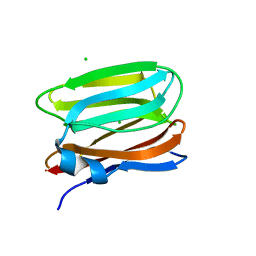
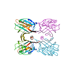
3VZE
DownloadVisualize
BU of 3vze by Molmil
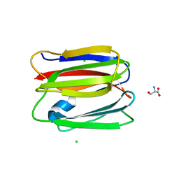
Crystal structure of human pancreatic secretory protein ZG16p with alpha1,3-mannobiose
Descriptor: CHLORIDE ION, Zymogen granule membrane protein 16, alpha-D-mannopyranose-(1-3)-alpha-D-mannopyranose
Authors:Kanagawa, M, Yamaguchi, Y.
Deposit date:2012-10-11
Release date:2013-10-16
Last modified:2023-11-08
Method:X-RAY DIFFRACTION (1.9 Å)
Cite:Structural Basis for Multiple Sugar Recognition of Jacalin-related Human ZG16p Lectin
J.Biol.Chem., 289, 2014
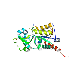
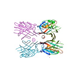
2ABK
DownloadVisualize
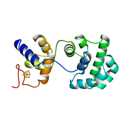
BU of 2abk by Molmil
REFINEMENT OF THE NATIVE STRUCTURE OF ENDONUCLEASE III TO A RESOLUTION OF 1.85 ANGSTROM
Descriptor: ENDONUCLEASE III, IRON/SULFUR CLUSTER
Authors:Thayer, M.M, Tainer, J.A.
Deposit date:1995-05-11
Release date:1995-10-15
Last modified:2024-02-14
Method:X-RAY DIFFRACTION (1.85 Å)
Cite:Novel DNA binding motifs in the DNA repair enzyme endonuclease III crystal structure.
EMBO J., 14, 1995
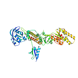
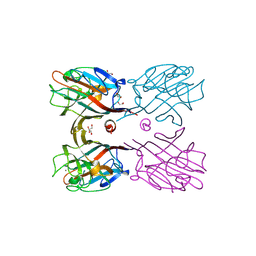
3U3D
DownloadVisualize
BU of 3u3d by Molmil
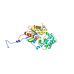
Plasmodium falciparum Sir2A preferentially hydrolyzes medium and long chain fatty acyl lysine
Descriptor: GLYCEROL, Transcriptional regulatory protein sir2 homologue, ZINC ION, ...
Authors:Zhou, Y, Hao, Q.
Deposit date:2011-10-05
Release date:2011-11-09
Last modified:2023-11-01
Method:X-RAY DIFFRACTION (2.4 Å)
Cite:Plasmodium falciparum Sir2A Preferentially Hydrolyzes Medium and Long Chain Fatty Acyl Lysine
Acs Chem.Biol., 2011
3TIX
DownloadVisualize
BU of 3tix by Molmil
Crystal structure of the Chp1-Tas3 complex core
Descriptor: CHLORIDE ION, Chromo domain-containing protein 1, POTASSIUM ION, ...
Authors:Schalch, T, Joshua-Tor, L.
Deposit date:2011-08-22
Release date:2011-11-16
Last modified:2024-02-28
Method:X-RAY DIFFRACTION (2.9001 Å)
Cite:The Chp1-Tas3 core is a multifunctional platform critical for gene silencing by RITS.
Nat.Struct.Mol.Biol., 18, 2011
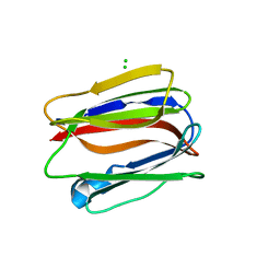
3VY6
DownloadVisualize
BU of 3vy6 by Molmil
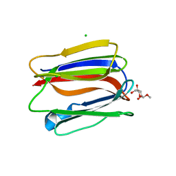
Crystal structure of human pancreatic secretory protein ZG16p with laminaribiose
Descriptor: CHLORIDE ION, Zymogen granule membrane protein 16, beta-D-glucopyranose-(1-3)-beta-D-glucopyranose
Authors:Kanagawa, M, Yamaguchi, Y.
Deposit date:2012-09-21
Release date:2013-09-25
Last modified:2023-11-08
Method:X-RAY DIFFRACTION (2 Å)
Cite:Structural Basis for Multiple Sugar Recognition of Jacalin-related Human ZG16p Lectin
J.Biol.Chem., 289, 2014
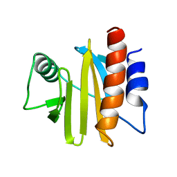
1A0K
DownloadVisualize
BU of 1a0k by Molmil
PROFILIN I FROM ARABIDOPSIS THALIANA
Descriptor: PROFILIN
Authors:Shigeta Junior, R, Huddler, D, Lindberg, U, Schutt, C.E.
Deposit date:1997-12-02
Release date:1998-03-18
Last modified:2024-05-22
Method:X-RAY DIFFRACTION (2.2 Å)
Cite:The crystal structure of a major allergen from plants.
Structure, 5, 1997
3VY7
DownloadVisualize
BU of 3vy7 by Molmil
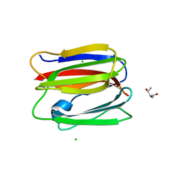
Crystal structure of human pancreatic secretory protein ZG16p with O-(alpha-D-mannosyl)-L-serine
Descriptor: CHLORIDE ION, SERINE, Zymogen granule membrane protein 16, ...
Authors:Kanagawa, M, Yamaguchi, Y.
Deposit date:2012-09-21
Release date:2013-09-25
Last modified:2023-11-08
Method:X-RAY DIFFRACTION (2.14 Å)
Cite:Structural Basis for Multiple Sugar Recognition of Jacalin-related Human ZG16p Lectin
J.Biol.Chem., 289, 2014
3VZF
DownloadVisualize
BU of 3vzf by Molmil
Crystal structure of human pancreatic secretory protein ZG16p with methyl alpha-D-mannopyranoside
Descriptor: CHLORIDE ION, Zymogen granule membrane protein 16, methyl alpha-D-mannopyranoside
Authors:Kanagawa, M, Yamaguchi, Y.
Deposit date:2012-10-11
Release date:2013-10-16
Last modified:2023-11-08
Method:X-RAY DIFFRACTION (2.8 Å)
Cite:Structural Basis for Multiple Sugar Recognition of Jacalin-related Human ZG16p Lectin
J.Biol.Chem., 289, 2014
3VZG
DownloadVisualize
BU of 3vzg by Molmil
Crystal structure of human pancreatic secretory protein ZG16p with O-(alpha-D-mannosyl)-L-threonine
Descriptor: CHLORIDE ION, THREONINE, Zymogen granule membrane protein 16, ...
Authors:Kanagawa, M, Yamaguchi, Y.
Deposit date:2012-10-11
Release date:2013-10-16
Last modified:2023-11-08
Method:X-RAY DIFFRACTION (2.7 Å)
Cite:Structural Basis for Multiple Sugar Recognition of Jacalin-related Human ZG16p Lectin
J.Biol.Chem., 289, 2014
3V00
DownloadVisualize
BU of 3v00 by Molmil
Studies of a constitutively active G-alpha subunit provide insights into the mechanism of G protein activation.
Descriptor: GUANOSINE-5'-DIPHOSPHATE, Guanine nucleotide-binding protein G(t) subunit alpha-1/ Guanine nucleotide-binding protein G(i) subunit alpha-1 chimeric protein
Authors:Singh, G, Cerione, R.A.
Deposit date:2011-12-07
Release date:2012-04-18
Last modified:2023-09-13
Method:X-RAY DIFFRACTION (2.9 Å)
Cite:A constitutively active G-alpha subunit provide insights into the mechanism of G protein activation
Biochemistry, 51, 2012
3WOG
DownloadVisualize
BU of 3wog by Molmil
Crystal structure plant lectin in complex with ligand
Descriptor: 2-acetamido-2-deoxy-beta-D-glucopyranose, 2-acetamido-2-deoxy-beta-D-glucopyranose-(1-2)-alpha-D-mannopyranose, CALCIUM ION, ...
Authors:Nagae, M, Yamaguchi, Y.
Deposit date:2013-12-26
Release date:2014-04-09
Last modified:2024-10-16
Method:X-RAY DIFFRACTION (2 Å)
Cite:Phytohemagglutinin from Phaseolus vulgaris (PHA-E) displays a novel glycan recognition mode using a common legume lectin fold
Glycobiology, 24, 2014
3WCR
DownloadVisualize
BU of 3wcr by Molmil
Crystal structure of plant lectin (ligand-free form)
Descriptor: 2-acetamido-2-deoxy-beta-D-glucopyranose, BROMIDE ION, Erythroagglutinin
Authors:Nagae, M, Yamaguchi, Y.
Deposit date:2013-05-31
Release date:2014-04-09
Last modified:2024-10-16
Method:X-RAY DIFFRACTION (2.45 Å)
Cite:Phytohemagglutinin from Phaseolus vulgaris (PHA-E) displays a novel glycan recognition mode using a common legume lectin fold
Glycobiology, 24, 2014
3WCS
DownloadVisualize
BU of 3wcs by Molmil
Crystal structure of plant lectin (ligand-bound form)
Descriptor: 1,2-ETHANEDIOL, 2-acetamido-2-deoxy-beta-D-glucopyranose, 2-acetamido-2-deoxy-beta-D-glucopyranose-(1-2)-alpha-D-mannopyranose, ...
Authors:Nagae, M, Yamaguchi, Y.
Deposit date:2013-05-31
Release date:2014-04-09
Last modified:2024-10-09
Method:X-RAY DIFFRACTION (1.75 Å)
Cite:Phytohemagglutinin from Phaseolus vulgaris (PHA-E) displays a novel glycan recognition mode using a common legume lectin fold
Glycobiology, 24, 2014

227111

건을2024-11-06부터공개중

PDB statisticsPDBj update infoContact PDBjnumon