1O3U
 
 | |
1O22
 | |
1O4V
 | |
1KQ4
 | |
1VKY
 | |
1VR0
 | |
1VJ2
 | |
1VKB
 | |
1VQ3
 | |
1VKM
 | |
1VLR
 | |
1VJ1
 | |
1VQR
 | |
1VR8
 | |
1VPZ
 | |
1VKH
 | |
1VK3
 | |
1VL4
 | |
1VLP
 | |
1VQ0
 | |
1VJO
 | |
1VR3
 | |
1VRM
 | |
1EZZ
 | |
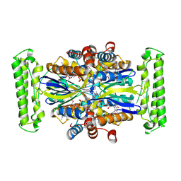
1I5O
 | | CRYSTAL STRUCTURE OF MUTANT R105A OF E. COLI ASPARTATE TRANSCARBAMOYLASE | | Descriptor: | ASPARTATE TRANSCARBAMOYLASE CATALYTIC CHAIN, ASPARTATE TRANSCARBAMOYLASE REGULATORY CHAIN, N-(PHOSPHONACETYL)-L-ASPARTIC ACID, ... | | Authors: | Macol, C.P, Tsuruta, H, Stec, B, Kantrowitz, E.R. | | Deposit date: | 2001-02-28 | | Release date: | 2001-05-02 | | Last modified: | 2023-08-09 | | Method: | X-RAY DIFFRACTION (2.8 Å) | | Cite: | Direct structural evidence for a concerted allosteric transition in Escherichia coli aspartate transcarbamoylase.
Nat.Struct.Biol., 8, 2001
|
|


