5VC2
 
 | |
5VMK
 | |
5VMB
 | |
3SDS
 | |
7JP4
 | |
7JFZ
 | |
7JFN
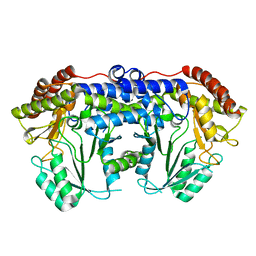
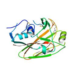
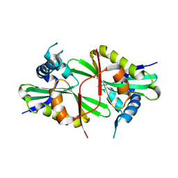
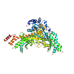
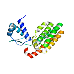
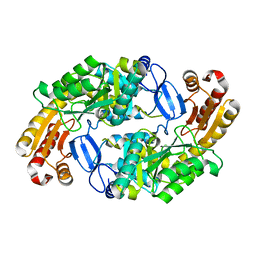
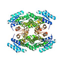
 | | Crystal Structure of Leucine-, isoleucine-, valine-, threonine-, and alanine-binding protein from Brucella ovis, closed conformation | | Descriptor: | 1,2-ETHANEDIOL, NITRATE ION, POTASSIUM ION, ... | | Authors: | Seattle Structural Genomics Center for Infectious Disease (SSGCID) | | Deposit date: | 2020-07-17 | | Release date: | 2020-07-29 | | Last modified: | 2023-10-18 | | Method: | X-RAY DIFFRACTION (1.7 Å) | | Cite: | Crystal Structure of Leucine-, isoleucine-, valine-, threonine-, and alanine-binding protein from Brucella ovis, closed conformation
to be published
|
|
8SJ9
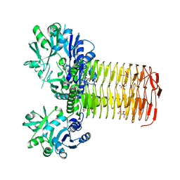
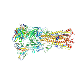
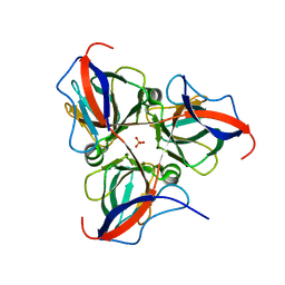
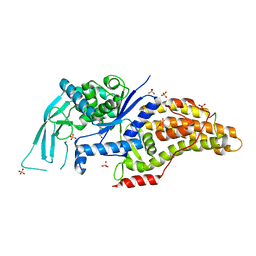
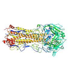 | | Crystal structure of the H1 hemagglutinin COBRA X6 | | Descriptor: | 1,2-ETHANEDIOL, 2-acetamido-2-deoxy-beta-D-glucopyranose, 2-acetamido-2-deoxy-beta-D-glucopyranose-(1-4)-2-acetamido-2-deoxy-beta-D-glucopyranose, ... | | Authors: | Dzimianski, J.V, DuBois, R.M. | | Deposit date: | 2023-04-17 | | Release date: | 2024-05-01 | | Method: | X-RAY DIFFRACTION (3.25 Å) | | Cite: | Structural basis for the expanded antibody breadth against X6, a broadly reactive H1 hemagglutinin vaccine
To Be Published
|
|
8V7O
 | |
7N1L
 | |
7N4D
 | |
7N6S
 | |
7N7S
 | |
7N56
 | |
7U4H
 | |
7U35
 | |
7U5Q
 | |
7U56
 | |
7U5Y
 | |
7U5F
 | |
7UG3
 | |
7ULH
 | |
7ULZ
 | |
7V0H
 | |
4TRR
 | |


