2B6M
 
 | |
2B3S
 | |
7QJF
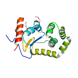
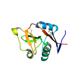
 | | Llp mutant C1G, lytic conversion lipoprotein of phage T5 | | 分子名称: | Lytic conversion lipoprotein | | 著者 | Degroux, S, Mestdach, E, Vives, C, Le Roy, A, Salmon, L, Herrman, T, Breyton, C. | | 登録日 | 2021-12-16 | | 公開日 | 2022-12-28 | | 実験手法 | SOLUTION NMR | | 主引用文献 | Llp mutant C1G, lytic conversion lipoprotein of phage T5
To Be Published
|
|
4AK8
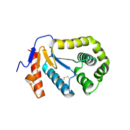
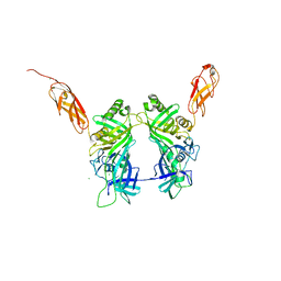
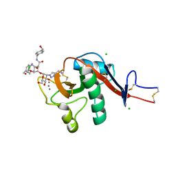
 | | Structure of F241L mutant of langerin carbohydrate recognition domain. | | 分子名称: | C-TYPE LECTIN DOMAIN FAMILY 4 MEMBER K, CALCIUM ION, CHLORIDE ION, ... | | 著者 | Chabrol, E, Thepaut, M, Dezutter-Dambuyant, C, Vives, C, Marcoux, J, Kahn, R, Valadeau-Guilemond, J, Vachette, P, Durand, D, Fieschi, F. | | 登録日 | 2012-02-22 | | 公開日 | 2013-04-03 | | 最終更新日 | 2023-12-20 | | 実験手法 | X-RAY DIFFRACTION (1.4 Å) | | 主引用文献 | Alteration of the Langerin Oligomerization State Affects Birbeck Granule Formation.
Biophys.J., 108, 2015
|
|
5NGJ
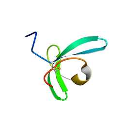
 | | Crystal structure of pb6, major tail tube protein of bacteriophage T5 | | 分子名称: | CHLORIDE ION, Tail tube protein | | 著者 | Arnaud, C.-A, Effantin, G, Vives, C, Engilberge, S, Bacia, M, Boulanger, P, Girard, E, Schoehn, G, Breyton, C. | | 登録日 | 2017-03-17 | | 公開日 | 2018-01-03 | | 最終更新日 | 2024-05-08 | | 実験手法 | X-RAY DIFFRACTION (2.2 Å) | | 主引用文献 | Bacteriophage T5 tail tube structure suggests a trigger mechanism for Siphoviridae DNA ejection.
Nat Commun, 8, 2017
|
|
3C22
 | |
1TI1
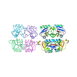
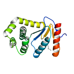 | | crystal structure of a mutant DsbA | | 分子名称: | DODECANE, Thiol:disulfide interchange protein dsbA | | 著者 | Kone, A, Serre, L. | | 登録日 | 2004-06-02 | | 公開日 | 2005-05-03 | | 最終更新日 | 2023-08-23 | | 実験手法 | X-RAY DIFFRACTION (2.6 Å) | | 主引用文献 | Intriguing conformation changes associated with the trans/cis isomerization of a prolyl residue in the active site of the DsbA C33A mutant.
J.Mol.Biol., 347, 2005
|
|
1U3A
 | | mutant DsbA | | 分子名称: | 3,6,9,12,15,18,21,24-OCTAOXAHEXACOSAN-1-OL, Thiol: disulfide interchange protein dsbA | | 著者 | Serre, L. | | 登録日 | 2004-07-21 | | 公開日 | 2005-05-03 | | 最終更新日 | 2023-08-23 | | 実験手法 | X-RAY DIFFRACTION (2 Å) | | 主引用文献 | Intriguing conformation changes associated with the trans/cis isomerization of a prolyl residue in the active site of the DsbA C33A mutant
J.Mol.Biol., 347, 2005
|
|
6GHV
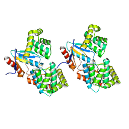
 | | Structure of a DC-SIGN CRD in complex with high affinity glycomimetic. | | 分子名称: | CALCIUM ION, CD209 antigen, CHLORIDE ION, ... | | 著者 | Thepaut, M, Achilli, S, Medve, L, Bernardi, A, Fieschi, F. | | 登録日 | 2018-05-09 | | 公開日 | 2019-09-11 | | 最終更新日 | 2024-01-17 | | 実験手法 | X-RAY DIFFRACTION (2.1 Å) | | 主引用文献 | Enhancing Potency and Selectivity of a DC-SIGN Glycomimetic Ligand by Fragment-Based Design: Structural Basis.
Chemistry, 25, 2019
|
|


