4S0W
 
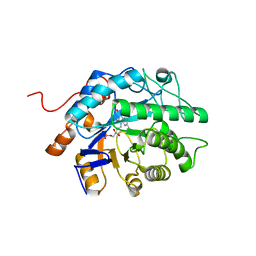
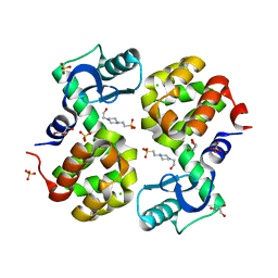 | | Wild type T4 lysozyme structure | | 分子名称: | 4-(2-HYDROXYETHYL)-1-PIPERAZINE ETHANESULFONIC ACID, CHLORIDE ION, GLYCEROL, ... | | 著者 | Snell, E.H, Snell, M.E. | | 登録日 | 2015-01-07 | | 公開日 | 2015-02-04 | | 最終更新日 | 2023-09-20 | | 実験手法 | X-RAY DIFFRACTION (2.117 Å) | | 主引用文献 | Wild type T4 lysozyme structure
To be Published
|
|
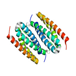
6VE1
 | |
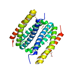
6P7J
 | |
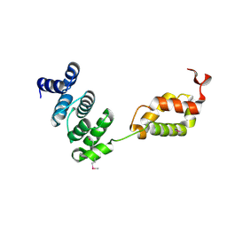
8FUH
 | |
8FVV
 | |
8FXD
 | |
3TL4
 | |
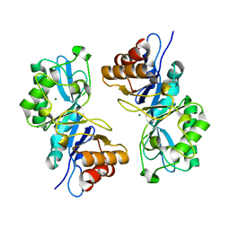
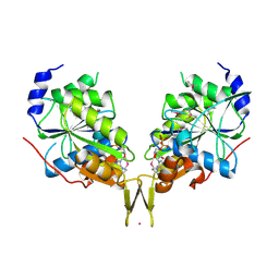
6NLR
 | | Crystal structure of the putative histidinol phosphatase hisK from Listeria monocytogenes with trinuclear metals determined by PIXE revealing sulphate ion in active site. Based on PIXE analysis and original date from 3DCP | | 分子名称: | CALCIUM ION, COBALT (II) ION, FE (III) ION, ... | | 著者 | Snell, E.H, Garman, E.F, Lowe, E.D. | | 登録日 | 2019-01-09 | | 公開日 | 2019-12-25 | | 最終更新日 | 2020-01-22 | | 実験手法 | X-RAY DIFFRACTION (2.1 Å) | | 主引用文献 | High-Throughput PIXE as an Essential Quantitative Assay for Accurate Metalloprotein Structural Analysis: Development and Application.
J.Am.Chem.Soc., 142, 2020
|
|
6OE2
 | |
6OBY
 | |
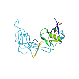
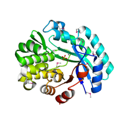
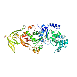
8FW1
 | | Gluconobacter Ene-Reductase (GluER) mutant - PagER | | 分子名称: | FLAVIN MONONUCLEOTIDE, N-ethylmaleimide reductase | | 著者 | Dahagam, S, Page, C, Patterson, M.G, Hyster, T.K. | | 登録日 | 2023-01-20 | | 公開日 | 2023-06-28 | | 最終更新日 | 2023-10-25 | | 実験手法 | X-RAY DIFFRACTION (1.5 Å) | | 主引用文献 | Regioselective Radical Alkylation of Arenes Using Evolved Photoenzymes.
J.Am.Chem.Soc., 145, 2023
|
|
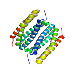
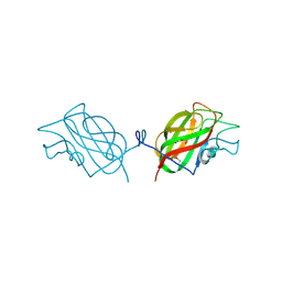
4H3S
 | | The Structure of Glutaminyl-tRNA Synthetase from Saccharomyces Cerevisiae | | 分子名称: | 2,3-DIHYDROXY-1,4-DITHIOBUTANE, ACETATE ION, BROMIDE ION, ... | | 著者 | Snell, E.H, Grant, T.D. | | 登録日 | 2012-09-14 | | 公開日 | 2013-04-24 | | 最終更新日 | 2023-09-20 | | 実験手法 | X-RAY DIFFRACTION (2.15 Å) | | 主引用文献 | The Structure of Yeast Glutaminyl-tRNA Synthetase and Modeling of Its Interaction with tRNA.
J.Mol.Biol., 425, 2013
|
|


