5I7Q
 
 | |
5I7P
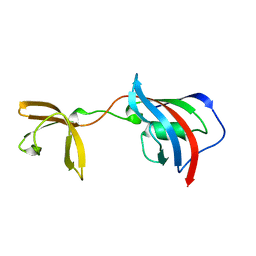
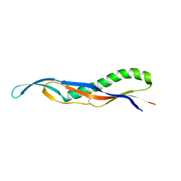
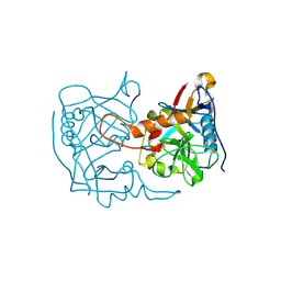
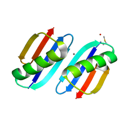
 | | Crystal structure of Fkbp12-IF(SlyD), a chimeric protein of human Fkbp12 and the insert in flap domain of Ecoli SlyD | | 分子名称: | Peptidyl-prolyl cis-trans isomerase FKBP1A,FKBP-type peptidyl-prolyl cis-trans isomerase SlyD,Peptidyl-prolyl cis-trans isomerase FKBP1A | | 著者 | Jakob, R.P, Knappe, T.A, Dobbek, H, Schmid, F.X. | | 登録日 | 2016-02-18 | | 公開日 | 2017-03-08 | | 最終更新日 | 2024-01-10 | | 実験手法 | X-RAY DIFFRACTION (2.002 Å) | | 主引用文献 | Structural and Functional Analysis of Chaperone Domain Insertion in Fkbp12
To Be Published
|
|
2X9B
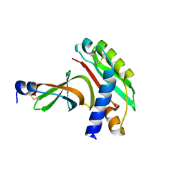
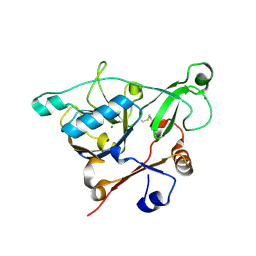
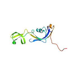
 | | The filamentous phages fd and IF1 use different infection mechanisms | | 分子名称: | ATTACHMENT PROTEIN G3P | | 著者 | Lorenz, S.H, Jakob, R.P, Weininger, U, Dobbek, H, Schmid, F.X. | | 登録日 | 2010-03-15 | | 公開日 | 2010-12-01 | | 最終更新日 | 2023-12-20 | | 実験手法 | X-RAY DIFFRACTION (2.92 Å) | | 主引用文献 | The Filamentous Phages Fd and If1 Use Different Mechanisms to Infect Escherichia Coli.
J.Mol.Biol., 405, 2011
|
|
2X9A
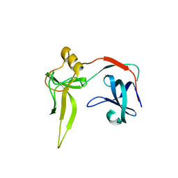
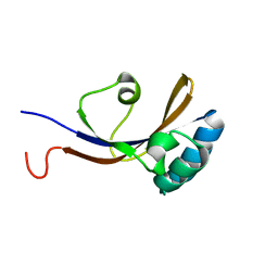
 | | crystal structure of g3p from phage IF1 in complex with its coreceptor, the C-terminal domain of TolA | | 分子名称: | ATTACHMENT PROTEIN G3P, MEMBRANE SPANNING PROTEIN, REQUIRED FOR OUTER MEMBRANE INTEGRITY | | 著者 | Lorenz, S.H, Jakob, R.P, Dobbek, H, Schmid, F.X. | | 登録日 | 2010-03-15 | | 公開日 | 2010-12-01 | | 最終更新日 | 2023-12-20 | | 実験手法 | X-RAY DIFFRACTION (2.47 Å) | | 主引用文献 | The Filamentous Phages Fd and If1 Use Different Mechanisms to Infect Escherichia Coli.
J.Mol.Biol., 405, 2011
|
|
3DGS
 | |
3DTM
 | |
4EO0
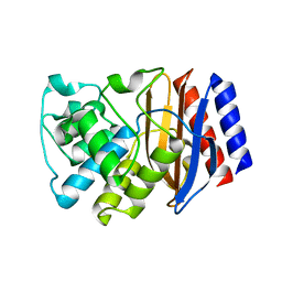
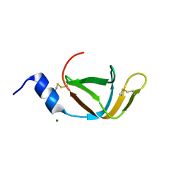
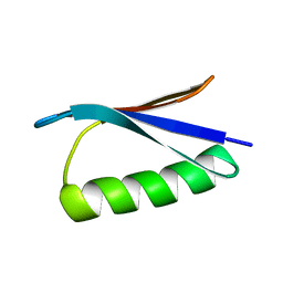
 | | crystal structure of the pilus binding domain of the filamentous phage IKe | | 分子名称: | Attachment protein G3P | | 著者 | Jakob, R.P, Geitner, A.J, Weininger, U, Balbach, J, Dobbek, H, Schmid, F.X. | | 登録日 | 2012-04-13 | | 公開日 | 2012-05-30 | | 最終更新日 | 2017-10-25 | | 実験手法 | X-RAY DIFFRACTION (1.61 Å) | | 主引用文献 | Structural and energetic basis of infection by the filamentous bacteriophage IKe.
Mol.Microbiol., 84, 2012
|
|
4EO1
 | | crystal structure of the TolA binding domain from the filamentous phage IKe | | 分子名称: | Attachment protein G3P, MAGNESIUM ION | | 著者 | Jakob, R.P, Geitner, A.J, Weininger, U, Balbach, J, Dobbek, H, Schmid, F.X. | | 登録日 | 2012-04-13 | | 公開日 | 2012-05-30 | | 最終更新日 | 2023-09-13 | | 実験手法 | X-RAY DIFFRACTION (1.8 Å) | | 主引用文献 | Structural and energetic basis of infection by the filamentous bacteriophage IKe.
Mol.Microbiol., 84, 2012
|
|
1C9O
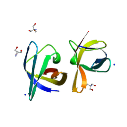 | | CRYSTAL STRUCTURE ANALYSIS OF THE BACILLUS CALDOLYTICUS COLD SHOCK PROTEIN BC-CSP | | 分子名称: | 2-AMINO-2-HYDROXYMETHYL-PROPANE-1,3-DIOL, COLD-SHOCK PROTEIN, SODIUM ION | | 著者 | Mueller, U, Perl, D, Schmid, F.X, Heinemann, U. | | 登録日 | 1999-08-03 | | 公開日 | 2000-04-02 | | 最終更新日 | 2024-02-07 | | 実験手法 | X-RAY DIFFRACTION (1.17 Å) | | 主引用文献 | Thermal stability and atomic-resolution crystal structure of the Bacillus caldolyticus cold shock protein.
J.Mol.Biol., 297, 2000
|
|
1HZ9
 | | BACILLUS CALDOLYTICUS COLD-SHOCK PROTEIN MUTANTS TO STUDY DETERMINANTS OF PROTEIN STABILITY | | 分子名称: | COLD SHOCK PROTEIN CSPB | | 著者 | Delbrueck, H, Mueller, U, Perl, D, Schmid, F.X, Heinemann, U. | | 登録日 | 2001-01-24 | | 公開日 | 2001-11-07 | | 最終更新日 | 2023-08-09 | | 実験手法 | X-RAY DIFFRACTION (1.8 Å) | | 主引用文献 | Crystal structures of mutant forms of the Bacillus caldolyticus cold shock protein differing in thermal stability.
J.Mol.Biol., 313, 2001
|
|
1HZA
 | | BACILLUS CALDOLYTICUS COLD-SHOCK PROTEIN MUTANTS TO STUDY DETERMINANTS OF PROTEIN STABILITY | | 分子名称: | COLD SHOCK PROTEIN CSPB | | 著者 | Delbrueck, H, Mueller, U, Perl, D, Schmid, F.X, Heinemann, U. | | 登録日 | 2001-01-24 | | 公開日 | 2001-11-07 | | 最終更新日 | 2023-08-09 | | 実験手法 | X-RAY DIFFRACTION (1.8 Å) | | 主引用文献 | Crystal structures of mutant forms of the Bacillus caldolyticus cold shock protein differing in thermal stability.
J.Mol.Biol., 313, 2001
|
|
1HZB
 | | BACILLUS CALDOLYTICUS COLD-SHOCK PROTEIN MUTANTS TO STUDY DETERMINANTS OF PROTEIN STABILITY | | 分子名称: | COLD SHOCK PROTEIN CSPB, SODIUM ION | | 著者 | Delbrueck, H, Mueller, U, Perl, D, Schmid, F.X, Heinemann, U. | | 登録日 | 2001-01-24 | | 公開日 | 2001-11-07 | | 最終更新日 | 2023-08-09 | | 実験手法 | X-RAY DIFFRACTION (1.28 Å) | | 主引用文献 | Crystal structures of mutant forms of the Bacillus caldolyticus cold shock protein differing in thermal stability.
J.Mol.Biol., 313, 2001
|
|
1HZC
 | | BACILLUS CALDOLYTICUS COLD-SHOCK PROTEIN MUTANTS TO STUDY DETERMINANTS OF PROTEIN STABILITY | | 分子名称: | COLD SHOCK PROTEIN CSPB, SODIUM ION | | 著者 | Delbrueck, H, Mueller, U, Perl, D, Schmid, F.X, Heinemann, U. | | 登録日 | 2001-01-24 | | 公開日 | 2001-11-07 | | 最終更新日 | 2023-08-09 | | 実験手法 | X-RAY DIFFRACTION (1.32 Å) | | 主引用文献 | Crystal structures of mutant forms of the Bacillus caldolyticus cold shock protein differing in thermal stability.
J.Mol.Biol., 313, 2001
|
|
1I5F
 | | BACILLUS CALDOLYTICUS COLD-SHOCK PROTEIN MUTANTS TO STUDY DETERMINANTS OF PROTEIN STABILITY | | 分子名称: | COLD-SHOCK PROTEIN CSPB, SODIUM ION | | 著者 | Delbrueck, H, Mueller, U, Perl, D, Schmid, F.X, Heinemann, U. | | 登録日 | 2001-02-27 | | 公開日 | 2001-11-07 | | 最終更新日 | 2023-08-09 | | 実験手法 | X-RAY DIFFRACTION (1.4 Å) | | 主引用文献 | Crystal structures of mutant forms of the Bacillus caldolyticus cold shock protein differing in thermal stability.
J.Mol.Biol., 313, 2001
|
|
2I5M
 | |
2I5L
 | |
4WO7
 | |
2ON8
 | |
5EX1
 | |
5EX2
 | |
2ONQ
 | |
3KNQ
 | |
3FIL
 | |
2K8I
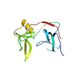
 | | Solution structure of E.Coli SlyD | | 分子名称: | Peptidyl-prolyl cis-trans isomerase | | 著者 | Weininger, U, Balbach, J. | | 登録日 | 2008-09-11 | | 公開日 | 2009-03-24 | | 最終更新日 | 2024-05-01 | | 実験手法 | SOLUTION NMR | | 主引用文献 | NMR solution structure of SlyD from Escherichia coli: spatial separation of prolyl isomerase and chaperone function.
J.Mol.Biol., 387, 2009
|
|
2KGJ
 | |


