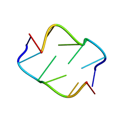
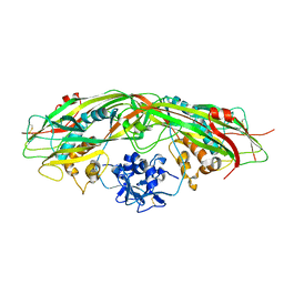
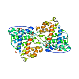
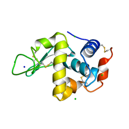
1LVY
 
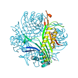
 | | PORCINE ELASTASE | | 分子名称: | CALCIUM ION, ELASTASE, SULFATE ION | | 著者 | Schiltz, M, Prange, T. | | 登録日 | 1996-07-20 | | 公開日 | 1997-01-27 | | 最終更新日 | 2023-08-09 | | 実験手法 | X-RAY DIFFRACTION (1.87 Å) | | 主引用文献 | High-pressure krypton gas and statistical heavy-atom refinement: a successful combination of tools for macromolecular structure determination.
Acta Crystallogr.,Sect.D, 53, 1997
|
|
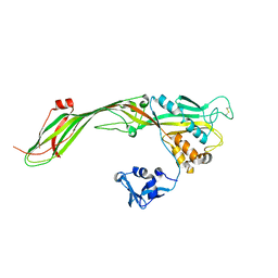
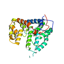
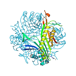
2OBZ
 | |
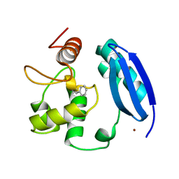
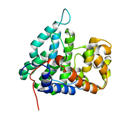
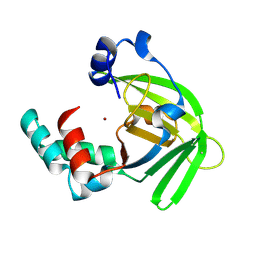
3KZY
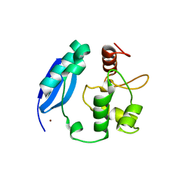 | | Crystal structure of SNAP-tag | | 分子名称: | Methylated-DNA--protein-cysteine methyltransferase, ZINC ION | | 著者 | Bannwarth, M, Schmitt, S, Pojer, F, Schiltz, M, Johnsson, K. | | 登録日 | 2009-12-09 | | 公開日 | 2010-12-15 | | 最終更新日 | 2023-11-08 | | 実験手法 | X-RAY DIFFRACTION (1.9 Å) | | 主引用文献 | SNAP-tag structure
To be Published
|
|
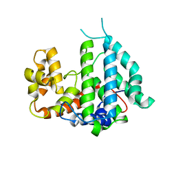
1C1M
 | | PORCINE ELASTASE UNDER XENON PRESSURE (8 BAR) | | 分子名称: | CALCIUM ION, PROTEIN (PORCINE ELASTASE), SULFATE ION, ... | | 著者 | Prange, T, Schiltz, M, Pernot, L, Colloc'h, N, Longhi, S, Bourguet, W, Fourme, R. | | 登録日 | 1999-07-22 | | 公開日 | 1999-07-28 | | 最終更新日 | 2018-02-28 | | 実験手法 | X-RAY DIFFRACTION (2.2 Å) | | 主引用文献 | Exploring hydrophobic sites in proteins with xenon or krypton.
Proteins, 30, 1998
|
|
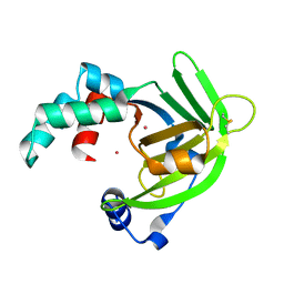
3KZZ
 | |
1C3L
 | | SUBTILISIN-CARLSBERG COMPLEXED WITH XENON (8 BAR) | | 分子名称: | CALCIUM ION, FORMIC ACID, SUBTILISIN-CARLSBERG, ... | | 著者 | Prange, T, Schiltz, M, Pernot, L, Colloc'h, N, Longhi, S. | | 登録日 | 1999-07-28 | | 公開日 | 1999-08-04 | | 最終更新日 | 2023-08-09 | | 実験手法 | X-RAY DIFFRACTION (2.16 Å) | | 主引用文献 | Exploring hydrophobic sites in proteins with xenon or krypton.
Proteins, 30, 1998
|
|
3L00
 | |
2CKK
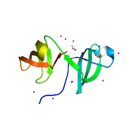 | | High resolution crystal structure of the human kin17 C-terminal domain containing a kow motif | | 分子名称: | ACETATE ION, IODIDE ION, KIN17 | | 著者 | le Maire, A, Schiltz, M, Pinon-Lataillade, G, Stura, E, Couprie, J, Gondry, M, Angulo-Mora, J, Zinn-Justin, S. | | 登録日 | 2006-04-20 | | 公開日 | 2006-10-04 | | 最終更新日 | 2024-05-08 | | 実験手法 | X-RAY DIFFRACTION (1.45 Å) | | 主引用文献 | A Tandem of SH3-Like Domains Participates in RNA Binding in Kin17, a Human Protein Activated in Response to Genotoxics.
J.Mol.Biol., 364, 2006
|
|
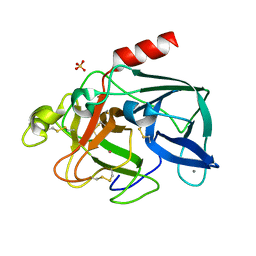
2C5Q
 | | Crystal structure of yeast YER010Cp | | 分子名称: | 1,2-ETHANEDIOL, RRAA-LIKE PROTEIN YER010C | | 著者 | Leulliot, N, Quevillon-Cheruel, S, Graille, M, Schiltz, M, Blondeau, K, Janin, J, van Tilbeurgh, H. | | 登録日 | 2005-10-31 | | 公開日 | 2005-11-14 | | 最終更新日 | 2011-07-13 | | 実験手法 | X-RAY DIFFRACTION (1.7 Å) | | 主引用文献 | Crystal Structure of Yeast Yer010Cp, a Knotable Member of the Rraa Protein Family.
Protein Sci., 14, 2005
|
|
3G4O
 | |
3G4N
 | |
1C10
 | | CRYSTAL STRUCTURE OF HEW LYSOZYME UNDER PRESSURE OF XENON (8 BAR) | | 分子名称: | CHLORIDE ION, PROTEIN (LYSOZYME), SODIUM ION, ... | | 著者 | Prange, T, Schiltz, M, Pernot, L, Colloc'h, N, Longhi, S, Bourguet, W, Fourme, R. | | 登録日 | 1999-07-16 | | 公開日 | 1999-07-22 | | 最終更新日 | 2023-08-09 | | 実験手法 | X-RAY DIFFRACTION (2.03 Å) | | 主引用文献 | Exploring hydrophobic sites in proteins with xenon or krypton.
Proteins, 30, 1998
|
|
3GNX
 | |
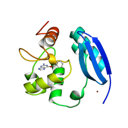
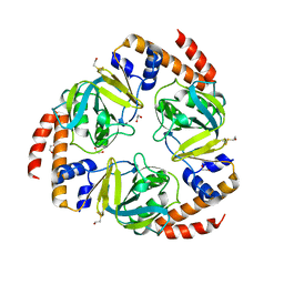
2IXP
 | | Crystal structure of the Pp2A phosphatase activator Ypa1 PTPA1 in complex with model substrate | | 分子名称: | CHLORIDE ION, SERINE/THREONINE-PROTEIN PHOSPHATASE 2A ACTIVATOR 1, SIN-ALA-ALA-PRO-LYS-NIT, ... | | 著者 | Leulliot, N, Vicentini, G, Jordens, J, Quevillon-Cheruel, S, Schiltz, M, Barford, D, Van Tilbeurgh, H, Goris, J. | | 登録日 | 2006-07-09 | | 公開日 | 2006-07-31 | | 最終更新日 | 2023-12-13 | | 実験手法 | X-RAY DIFFRACTION (2.8 Å) | | 主引用文献 | Crystal structure of the PP2A phosphatase activator: implications for its PP2A-specific PPIase activity.
Mol. Cell, 23, 2006
|
|
2IXO
 | | CRYSTAL STRUCTURE OF THE PP2A PHOSPHATASE ACTIVATOR Ypa1 PTPA1 | | 分子名称: | SERINE/THREONINE-PROTEIN PHOSPHATASE 2A ACTIVATOR 1 | | 著者 | Leulliot, N, Vicentini, G, Jordens, J, Quevillon-Cheruel, S, Schiltz, M, Barford, D, Van Tilbeurgh, H, Goris, J. | | 登録日 | 2006-07-09 | | 公開日 | 2006-07-31 | | 最終更新日 | 2024-05-08 | | 実験手法 | X-RAY DIFFRACTION (2.6 Å) | | 主引用文献 | Crystal Structure of the Pp2A Phosphatase Activator: Implications for its Pp2A-Specific Ppiase Activity.
Mol.Cell, 23, 2006
|
|
2IXM
 | | Structure of human PTPA | | 分子名称: | SERINE/THREONINE-PROTEIN PHOSPHATASE 2A REGULATORY SUBUNIT B' | | 著者 | Leulliot, N, Vicentini, G, Jordens, J, Quevillon-Cheruel, S, Schiltz, M, Barford, D, Van Tilbeurgh, H, Goris, J. | | 登録日 | 2006-07-09 | | 公開日 | 2006-07-11 | | 最終更新日 | 2024-05-08 | | 実験手法 | X-RAY DIFFRACTION (1.5 Å) | | 主引用文献 | Crystal Structure of the Pp2A Phosphatase Activator: Implications for its Pp2A-Specific Ppiase Activity
Mol.Cell, 23, 2006
|
|
2IXN
 | | CRYSTAL STRUCTURE OF THE PP2A PHOSPHATASE ACTIVATOR Ypa2 PTPA2 | | 分子名称: | SERINE/THREONINE-PROTEIN PHOSPHATASE 2A ACTIVATOR 2 | | 著者 | Leulliot, N, Vicentini, G, Jordens, J, Quevillon-Cheruel, S, Schiltz, M, Barford, D, Van Tilbeurgh, H, Goris, J. | | 登録日 | 2006-07-09 | | 公開日 | 2006-07-31 | | 最終更新日 | 2024-05-08 | | 実験手法 | X-RAY DIFFRACTION (2.8 Å) | | 主引用文献 | Crystal Structure of the Pp2A Phosphatase Activator: Implications for its Pp2A-Specific Ppiase Activity
Mol.Cell, 23, 2006
|
|
1QTK
 | | CRYSTAL STRUCTURE OF HEW LYSOZYME UNDER PRESSURE OF KRYPTON (55 BAR) | | 分子名称: | CHLORIDE ION, KRYPTON, LYSOZYME, ... | | 著者 | Prange, T, Schiltz, M, Pernot, L, Colloc'h, N, Longhi, S, Bourguet, W, Fourme, R. | | 登録日 | 1999-06-28 | | 公開日 | 1999-07-06 | | 最終更新日 | 2011-07-13 | | 実験手法 | X-RAY DIFFRACTION (2.03 Å) | | 主引用文献 | Exploring hydrophobic sites in proteins with xenon or krypton.
Proteins, 30, 1998
|
|
3L8W
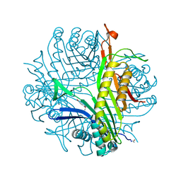 | | Urate oxidase from aspergillus flavus complexed with xanthin | | 分子名称: | SODIUM ION, Uricase, XANTHINE, ... | | 著者 | Prange, T, Gabison, L, Colloc'h, N, Chiadmi, M. | | 登録日 | 2010-01-04 | | 公開日 | 2010-06-02 | | 最終更新日 | 2023-11-01 | | 実験手法 | X-RAY DIFFRACTION (1 Å) | | 主引用文献 | Near-atomic resolution structures of urate oxidase complexed with its substrate and analogues: the protonation state of the ligand.
Acta Crystallogr.,Sect.D, 66, 2010
|
|
3CKU
 | |
1R56
 | |
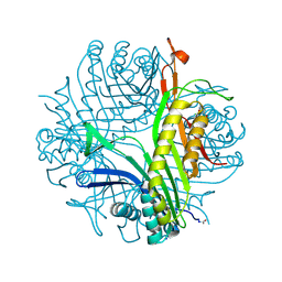
1R4S
 | | URATE OXIDASE FROM ASPERGILLUS FLAVUS COMPLEXED WITH ITS INHIBITOR 9-METHYL URIC ACID | | 分子名称: | 9-METHYL URIC ACID, CYSTEINE, Uricase | | 著者 | Retailleau, P, Colloc'h, N, Prange, T. | | 登録日 | 2003-10-08 | | 公開日 | 2004-03-02 | | 最終更新日 | 2023-12-13 | | 実験手法 | X-RAY DIFFRACTION (1.8 Å) | | 主引用文献 | Complexed and ligand-free high-resolution structures of urate oxidase (Uox) from Aspergillus flavus: a reassignment of the active-site binding mode.
Acta Crystallogr.,Sect.D, 60, 2004
|
|
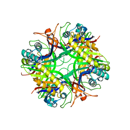
1R51
 | | URATE OXIDASE FROM ASPERGILLUS FLAVUS COMPLEXED WITH ITS INHIBITOR 8-AZAXANTHIN | | 分子名称: | 8-AZAXANTHINE, CYSTEINE, Uricase | | 著者 | Prange, T, Retailleau, P, Colloc'h, N. | | 登録日 | 2003-10-09 | | 公開日 | 2004-03-02 | | 最終更新日 | 2023-12-13 | | 実験手法 | X-RAY DIFFRACTION (1.75 Å) | | 主引用文献 | Complexed and ligand-free high-resolution structures of urate oxidase (Uox) from Aspergillus flavus: a reassignment of the active-site binding mode.
Acta Crystallogr.,Sect.D, 60, 2004
|
|
1OEJ
 | | YodA from Escherichia coli crystallised with no added ions | | 分子名称: | HYPOTHETICAL PROTEIN YODA, NICKEL (II) ION | | 著者 | David, G, Blondeau, K, Renouard, M, Penel, S, Lewit-Bentley, A. | | 登録日 | 2003-03-27 | | 公開日 | 2003-08-15 | | 最終更新日 | 2023-12-13 | | 実験手法 | X-RAY DIFFRACTION (1.81 Å) | | 主引用文献 | Yoda from Escherichia Coli is a Metal-Binding, Lipocalin-Like Protein
J.Biol.Chem., 278, 2003
|
|
1OEE
 | | YodA from Escherichia coli crystallised with cadmium ions | | 分子名称: | CADMIUM ION, HYPOTHETICAL PROTEIN YODA | | 著者 | David, G, Blondeau, K, Renouard, M, Penel, S, Lewit-Bentley, A. | | 登録日 | 2003-03-27 | | 公開日 | 2003-08-15 | | 最終更新日 | 2024-05-01 | | 実験手法 | X-RAY DIFFRACTION (2.1 Å) | | 主引用文献 | Yoda from Escherichia Coli is a Metal-Binding, Lipocalin-Like Protein
J.Biol.Chem., 278, 2003
|
|


