2LA0
 
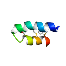 | | Trn- peptide of the two-component bacteriocin Thuricin CD | | 分子名称: | Uncharacterized protein | | 著者 | Sit, C.S, Mckay, R.T, Hill, C, Ross, R.P, Vederas, J.C. | | 登録日 | 2011-02-27 | | 公開日 | 2012-01-11 | | 最終更新日 | 2018-08-22 | | 実験手法 | SOLUTION NMR | | 主引用文献 | The 3D structure of thuricin CD, a two-component bacteriocin with cysteine sulfur to alpha-carbon cross-links.
J.Am.Chem.Soc., 133, 2011
|
|
2L9X
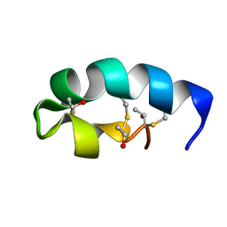 | | Trn- peptide of the two-component bacteriocin Thuricin CD | | 分子名称: | Uncharacterized protein | | 著者 | Sit, C.S, Mckay, R.T, Hill, C, Ross, R.P, Vederas, J.C. | | 登録日 | 2011-02-25 | | 公開日 | 2012-01-11 | | 最終更新日 | 2018-08-22 | | 実験手法 | SOLUTION NMR | | 主引用文献 | The 3D structure of thuricin CD, a two-component bacteriocin with cysteine sulfur to alpha-carbon cross-links.
J.Am.Chem.Soc., 133, 2011
|
|
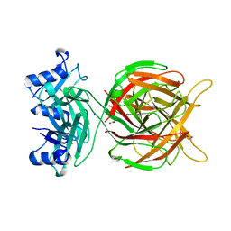
4CT3
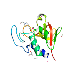 | | Methylmercury chloride derivative structure of the lytic CHAPK domain of the endolysin LysK from Staphylococcus aureus bacteriophage K | | 分子名称: | 4-(2-HYDROXYETHYL)-1-PIPERAZINE ETHANESULFONIC ACID, CALCIUM ION, CHLORIDE ION, ... | | 著者 | Sanz-Gaitero, M, Keary, R, Garcia-Doval, C, Coffey, A, van Raaij, M.J. | | 登録日 | 2014-03-12 | | 公開日 | 2014-08-06 | | 最終更新日 | 2024-06-26 | | 実験手法 | X-RAY DIFFRACTION (1.69 Å) | | 主引用文献 | Crystal structure of the lytic CHAP(K) domain of the endolysin LysK from Staphylococcus aureus bacteriophage K.
Virol. J., 11, 2014
|
|
4CSH
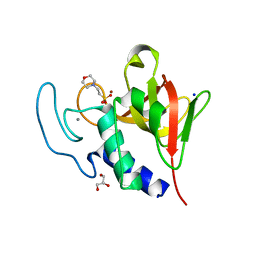 | | Native structure of the lytic CHAPK domain of the endolysin LysK from Staphylococcus aureus bacteriophage K | | 分子名称: | 2-(N-MORPHOLINO)-ETHANESULFONIC ACID, CALCIUM ION, GLYCEROL, ... | | 著者 | Sanz-Gaitero, M, Keary, R, Garcia-Doval, C, Coffey, A, van Raaij, M.J. | | 登録日 | 2014-03-07 | | 公開日 | 2014-08-06 | | 最終更新日 | 2023-12-20 | | 実験手法 | X-RAY DIFFRACTION (1.79 Å) | | 主引用文献 | Crystal structure of the lytic CHAP(K) domain of the endolysin LysK from Staphylococcus aureus bacteriophage K.
Virol. J., 11, 2014
|
|
5M9F
 | |
1NPX
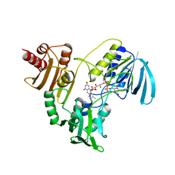 | | STRUCTURE OF NADH PEROXIDASE FROM STREPTOCOCCUS FAECALIS 10C1 REFINED AT 2.16 ANGSTROMS RESOLUTION | | 分子名称: | FLAVIN-ADENINE DINUCLEOTIDE, NADH PEROXIDASE | | 著者 | Stehle, T, Ahmed, S.A, Claiborne, A, Schulz, G.E. | | 登録日 | 1991-08-02 | | 公開日 | 1994-01-31 | | 最終更新日 | 2024-06-05 | | 実験手法 | X-RAY DIFFRACTION (2.16 Å) | | 主引用文献 | Structure of NADH peroxidase from Streptococcus faecalis 10C1 refined at 2.16 A resolution.
J.Mol.Biol., 221, 1991
|
|


