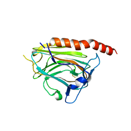
2JG9
 
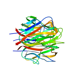 | | Crystallographic structure of human C1q globular heads (P1) | | 分子名称: | CALCIUM ION, Complement C1q subcomponent subunit A, Complement C1q subcomponent subunit B, ... | | 著者 | Paidassi, H, Tacnet-Delorme, P, Garlatti, V, Darnault, C, Ghebrehiwet, B, Gaboriaud, C, Arlaud, G.J, Frachet, P. | | 登録日 | 2007-02-09 | | 公開日 | 2008-02-19 | | 最終更新日 | 2023-12-13 | | 実験手法 | X-RAY DIFFRACTION (1.9 Å) | | 主引用文献 | C1Q Binds Phosphatidylserine and Likely Acts as a Multiligand-Bridging Molecule in Apoptotic Cell Recognition.
J.Immunol., 180, 2008
|
|
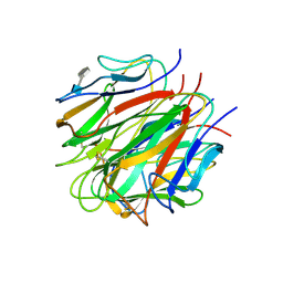
2JG8
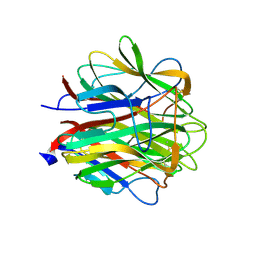 | | Crystallographic structure of human C1q globular heads complexed to phosphatidyl-serine | | 分子名称: | 2-acetamido-2-deoxy-beta-D-glucopyranose, CALCIUM ION, Complement C1q subcomponent subunit A, ... | | 著者 | Paidassi, H, Tacnet-Delorme, P, Garlatti, V, Darnault, C, Ghebrehiwet, B, Gaboriaud, C, Arlaud, G.J, Frachet, P. | | 登録日 | 2007-02-09 | | 公開日 | 2008-02-19 | | 最終更新日 | 2023-12-13 | | 実験手法 | X-RAY DIFFRACTION (2.05 Å) | | 主引用文献 | C1Q Binds Phosphatidylserine and Likely Acts as a Multiligand-Bridging Molecule in Apoptotic Cell Recognition.
J.Immunol., 180, 2008
|
|
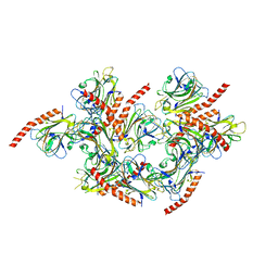
8BV0
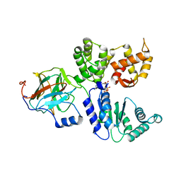 | | Binary complex between the NB-ARC domain from the Tomato immune receptor NRC1 and the SPRY domain-containing effector SS15 from the potato cyst nematode | | 分子名称: | ADENOSINE-5'-DIPHOSPHATE, NRC1, Truncated secreted SPRY domain-containing protein 15 (Fragment) | | 著者 | Contreras, M.P, Pai, H, Muniyandi, S, Toghani, A, Lawson, D.M, Tumtas, Y, Duggan, C, Yuen, E.L.H, Stevenson, C.E.M, Harant, A, Wu, C.H, Bozkurt, T.O, Kamoun, S, Derevnina, L. | | 登録日 | 2022-12-01 | | 公開日 | 2022-12-21 | | 最終更新日 | 2024-06-19 | | 実験手法 | X-RAY DIFFRACTION (4.5 Å) | | 主引用文献 | Resurrection of plant disease resistance proteins via helper NLR bioengineering.
Sci Adv, 9, 2023
|
|
4ACJ
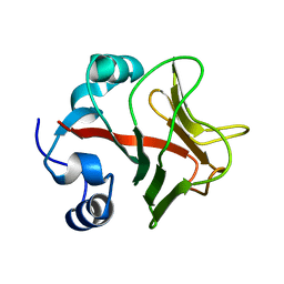 | | Crystal structure of the TLDC domain of Oxidation resistance protein 2 from zebrafish | | 分子名称: | WU:FB25H12 PROTEIN, | | 著者 | Blaise, M, B Alsarraf, H.M.A, Wong, J.E.M.M, Midtgaard, S.R, Laroche, F, Schack, L, Spaink, H, Stougaard, J, Thirup, S. | | 登録日 | 2011-12-15 | | 公開日 | 2012-02-08 | | 最終更新日 | 2024-05-08 | | 実験手法 | X-RAY DIFFRACTION (0.97 Å) | | 主引用文献 | Crystal Structure of the Tldc Domain of Oxidation Resistance Protein 2 from Zebrafish.
Proteins, 80, 2012
|
|
3POS
 | | Crystal structure of the globular domain of human calreticulin | | 分子名称: | CALCIUM ION, Calreticulin | | 著者 | Chouquet, A, Paidassi, H, Ling, W.-L, Frachet, P, Houen, G, Arlaud, G.J, Gaboriaud, C. | | 登録日 | 2010-11-23 | | 公開日 | 2011-03-09 | | 最終更新日 | 2017-08-09 | | 実験手法 | X-RAY DIFFRACTION (1.65 Å) | | 主引用文献 | X-ray structure of the human calreticulin globular domain reveals a Peptide-binding area and suggests a multi-molecular mechanism
Plos One, 6, 2011
|
|
2WNU
 | | Complex between c1q globular heads and heparan sulfate | | 分子名称: | 2-O-sulfo-beta-L-altropyranuronic acid-(1-4)-2-deoxy-6-O-sulfo-2-(sulfoamino)-alpha-D-glucopyranose, 2-acetamido-2-deoxy-beta-D-glucopyranose, COMPLEMENT C1Q SUBCOMPONENT SUBUNIT A, ... | | 著者 | Garlatti, V, Chouquet, A, Lunardi, T, Thielens, N.M, Arlaud, G.J, Gaboriaud, C. | | 登録日 | 2009-07-20 | | 公開日 | 2010-05-26 | | 最終更新日 | 2023-12-13 | | 実験手法 | X-RAY DIFFRACTION (2.3 Å) | | 主引用文献 | Cutting Edge: C1Q Binds Deoxyribose and Heparan Sulfate Through Neighboring Sites of its Recognition Domain.
J.Immunol., 185, 2010
|
|
2WNV
 | | Complex between C1q globular heads and deoxyribose | | 分子名称: | 2-acetamido-2-deoxy-beta-D-glucopyranose, 2-deoxy-beta-D-erythro-pentofuranose, CALCIUM ION, ... | | 著者 | Garlatti, V, Chouquet, A, Lunardi, T, Thielens, N.M, Arlaud, G.J, Gaboriaud, C. | | 登録日 | 2009-07-20 | | 公開日 | 2010-05-26 | | 最終更新日 | 2023-12-13 | | 実験手法 | X-RAY DIFFRACTION (1.25 Å) | | 主引用文献 | Cutting Edge: C1Q Binds Deoxyribose and Heparan Sulfate Through Neighboring Sites of its Recognition Domain.
J.Immunol., 185, 2010
|
|
5LK5
 | |
3POW
 | |


