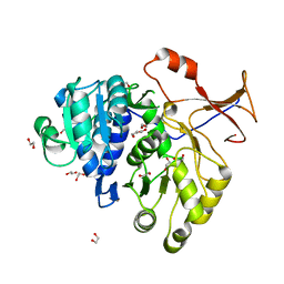
3DC0
 
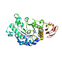 | | Crystal structure of native alpha-amylase from Bacillus sp. KR-8104 | | 分子名称: | CALCIUM ION, alpha-amylase | | 著者 | Alikhajeh, J, Khajeh, K, Ranjbar, B, Naderi-Manesh, M, Naderi-Manesh, H, Chen, C.J. | | 登録日 | 2008-06-03 | | 公開日 | 2008-06-17 | | 最終更新日 | 2023-11-01 | | 実験手法 | X-RAY DIFFRACTION (2.78 Å) | | 主引用文献 | Crystal structure of native alpha-amylase from Bacillus sp. KR-8104
to be published
|
|
1W6B
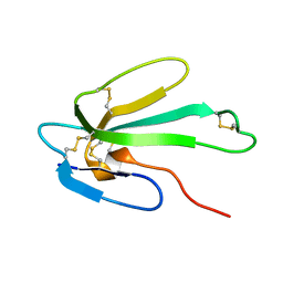 | | Solution NMR Structure of a Long Neurotoxin from the Venom of the Asian Cobra, 20 Structures | | 分子名称: | LONG NEUROTOXIN 1 | | 著者 | Talebzadeh-Farooji, M, Amininasab, M, Elmi, M.M, Naderi-Manesh, H, Sarbolouki, M.N. | | 登録日 | 2004-08-17 | | 公開日 | 2004-12-22 | | 最終更新日 | 2018-05-09 | | 実験手法 | SOLUTION NMR | | 主引用文献 | Solution structure of long neurotoxin NTX-1 from the venom of Naja naja oxiana by 2D-NMR spectroscopy.
Eur. J. Biochem., 271, 2004
|
|
2XW6
 | | The Crystal Structure of Methylglyoxal Synthase from Thermus sp. GH5 Bound to Phosphate Ion. | | 分子名称: | METHYLGLYOXAL SYNTHASE, PHOSPHATE ION | | 著者 | Shahsavar, A, Erfani Moghaddam, M, Antonyuk, S.V, Khajeh, K, Naderi-Manesh, H. | | 登録日 | 2010-11-01 | | 公開日 | 2011-11-09 | | 最終更新日 | 2023-12-20 | | 実験手法 | X-RAY DIFFRACTION (1.08 Å) | | 主引用文献 | Atomic Resolution Structure of Methylglyoxal Synthase from Thermus Sp. Gh5 Bound to Phosphate: Insights Into the Distinctive Effects of Phosphate on the Enzyme Structure
To be Published
|
|
2X8W
 | | The Crystal Structure of Methylglyoxal Synthase from Thermus sp. GH5 Bound to Malonate. | | 分子名称: | MALONATE ION, METHYLGLYOXAL SYNTHASE | | 著者 | Shahsavar, A, Erfani Moghaddam, M, Antonyuk, S.V, Khajeh, K, Naderi-Manesh, H. | | 登録日 | 2010-03-13 | | 公開日 | 2011-03-23 | | 最終更新日 | 2023-12-20 | | 実験手法 | X-RAY DIFFRACTION (1.95 Å) | | 主引用文献 | Crystal Structures of Methylglyoxal Synthase from Thermus Sp.Gh5 in the Open and Closed Conformational States Provide Insight Into the Mechanism of Allosteric Regulation
To be Published
|
|
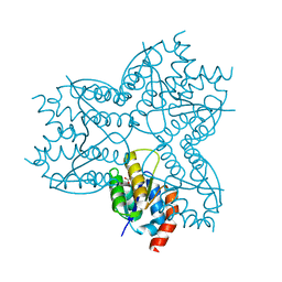
4M46
 | |
3QYA
 | | Crystal structure of a red-emitter mutant of Lampyris turkestanicus luciferase | | 分子名称: | 1,2-ETHANEDIOL, DI(HYDROXYETHYL)ETHER, GLYCEROL, ... | | 著者 | Kheirabadi, M, Gohlke, U, Hossein Khani, S, Heinemann, U, Naderi-Manesh, H. | | 登録日 | 2011-03-03 | | 公開日 | 2012-03-07 | | 最終更新日 | 2023-09-13 | | 実験手法 | X-RAY DIFFRACTION (2.13 Å) | | 主引用文献 | Crystal structure of native and a mutant of Lampyris turkestanicus luciferase implicate in bioluminescence color shift.
Biochim.Biophys.Acta, 1834, 2013
|
|
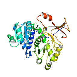
3BH4
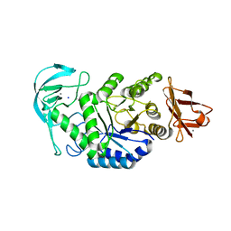 | | High resolution crystal structure of Bacillus amyloliquefaciens alpha-amylase | | 分子名称: | Alpha-amylase, CALCIUM ION, SODIUM ION | | 著者 | Alikhajeh, J, Khajeh, K, Ranjbar, B, Naderi-Manesh, H, Lin, Y.H, Liu, M.Y, Chen, C.J. | | 登録日 | 2007-11-28 | | 公開日 | 2008-12-09 | | 最終更新日 | 2023-11-01 | | 実験手法 | X-RAY DIFFRACTION (1.4 Å) | | 主引用文献 | Structure of Bacillus amyloliquefaciens alpha-amylase at high resolution: implications for thermal stability.
Acta Crystallogr.,Sect.F, 66, 2010
|
|
5VP3
 | | Light-sensitive photoprotein | | 分子名称: | CADMIUM ION, Mnemiopsin 1, NICKEL (II) ION, ... | | 著者 | Gorman, M.A, Parker, M.W. | | 登録日 | 2017-05-04 | | 公開日 | 2018-05-09 | | 最終更新日 | 2023-10-04 | | 実験手法 | X-RAY DIFFRACTION (2.151 Å) | | 主引用文献 | Reaction mechanism of the bioluminescent protein mnemiopsin1 revealed by X-ray crystallography and QM/MM simulations.
J. Biol. Chem., 294, 2019
|
|


