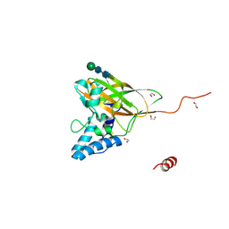
6VVG
 
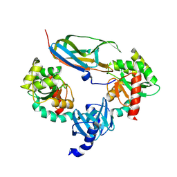
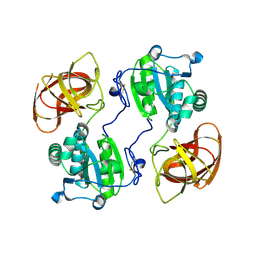
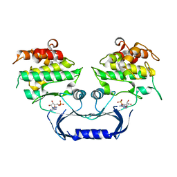 | | Structure of the Cydia pomonella Granulovirus kinase, PK-1 | | 分子名称: | ADENOSINE MONOPHOSPHATE, Arginine kinase | | 著者 | Oliver, M.R, Horne, C.R, Keown, J.R, Murphy, J.M, Metcalf, P. | | 登録日 | 2020-02-17 | | 公開日 | 2021-02-03 | | 最終更新日 | 2023-10-11 | | 実験手法 | X-RAY DIFFRACTION (2.01 Å) | | 主引用文献 | Granulovirus PK-1 kinase activity relies on a side-to-side dimerization mode centered on the regulatory alpha C helix.
Nat Commun, 12, 2021
|
|
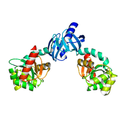
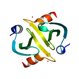
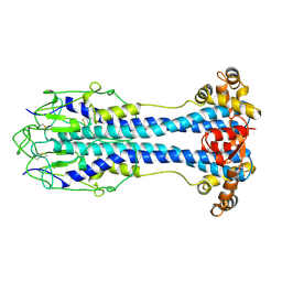
1EEJ
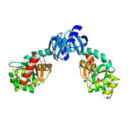 | | CRYSTAL STRUCTURE OF THE PROTEIN DISULFIDE BOND ISOMERASE, DSBC, FROM ESCHERICHIA COLI | | 分子名称: | 2-(N-MORPHOLINO)-ETHANESULFONIC ACID, THIOL:DISULFIDE INTERCHANGE PROTEIN | | 著者 | McCarthy, A.A, Haebel, P.W, Torronen, A, Rybin, V, Baker, E.N, Metcalf, P. | | 登録日 | 2000-01-31 | | 公開日 | 2000-08-03 | | 最終更新日 | 2011-07-13 | | 実験手法 | X-RAY DIFFRACTION (1.9 Å) | | 主引用文献 | Crystal structure of the protein disulfide bond isomerase, DsbC, from Escherichia coli.
Nat.Struct.Biol., 7, 2000
|
|
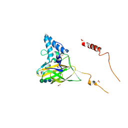
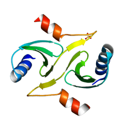
1G0T
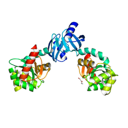 | | DSBC MUTANT C101S | | 分子名称: | DI(HYDROXYETHYL)ETHER, THIOL:DISULFIDE INTERCHANGE PROTEIN DSBC | | 著者 | Haebel, P.W, Metcalf, P. | | 登録日 | 2000-10-09 | | 公開日 | 2003-02-04 | | 最終更新日 | 2021-11-03 | | 実験手法 | X-RAY DIFFRACTION (2.6 Å) | | 主引用文献 | Crystal structure of the protein disulfide bond isomerase, DsbC, from Escherichia coli
Embo J., 21, 2002
|
|
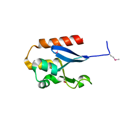
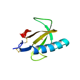
1LML
 | | LEISHMANOLYSIN | | 分子名称: | LEISHMANOLYSIN, ZINC ION | | 著者 | Schlagenhauf, E, Etges, R, Metcalf, P. | | 登録日 | 1997-03-13 | | 公開日 | 1997-09-17 | | 最終更新日 | 2011-07-13 | | 実験手法 | X-RAY DIFFRACTION (1.86 Å) | | 主引用文献 | The crystal structure of the Leishmania major surface proteinase leishmanolysin (gp63).
Structure, 6, 1998
|
|
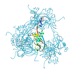
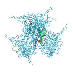
5G0Z
 | | Structure of native granulovirus polyhedrin determined using an X-ray free-electron laser | | 分子名称: | GRANULIN | | 著者 | Gati, C, Bunker, R.D, Oberthur, D, Metcalf, P, Henry, C. | | 登録日 | 2016-03-23 | | 公開日 | 2017-02-22 | | 最終更新日 | 2024-01-10 | | 実験手法 | X-RAY DIFFRACTION (2.001 Å) | | 主引用文献 | Atomic structure of granulin determined from native nanocrystalline granulovirus using an X-ray free-electron laser.
Proc. Natl. Acad. Sci. U.S.A., 114, 2017
|
|
5G3X
 | |
3JW6
 | |
3JVB
 | |
5CYY
 | |
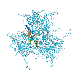
2OH6
 | | The Crystal Structure of Recombinant Cypovirus Polyhedra | | 分子名称: | ADENOSINE-5'-TRIPHOSPHATE, CHLORIDE ION, CYTIDINE-5'-TRIPHOSPHATE, ... | | 著者 | Coulibaly, F, Chiu, E, Ikeda, K, Gutmann, S, Haebel, P.W, Schulze-Briese, C, Mori, H, Metcalf, P. | | 登録日 | 2007-01-09 | | 公開日 | 2007-03-06 | | 最終更新日 | 2023-12-27 | | 実験手法 | X-RAY DIFFRACTION (2.1 Å) | | 主引用文献 | The molecular organization of cypovirus polyhedra.
Nature, 446, 2007
|
|
2OH5
 | | The Crystal Structure of Infectious Cypovirus Polyhedra | | 分子名称: | ADENOSINE-5'-TRIPHOSPHATE, CHLORIDE ION, CYTIDINE-5'-TRIPHOSPHATE, ... | | 著者 | Coulibaly, F, Chiu, E, Ikeda, K, Gutmann, S, Haebel, P.W, Schulze-Briese, C, Mori, H, Metcalf, P. | | 登録日 | 2007-01-09 | | 公開日 | 2007-03-06 | | 最終更新日 | 2023-12-27 | | 実験手法 | X-RAY DIFFRACTION (1.98 Å) | | 主引用文献 | The molecular organization of cypovirus polyhedra.
Nature, 446, 2007
|
|
2OH7
 | | The Crystal Structure of Cypovirus Polyhedra containing the Human ZIP-kinase | | 分子名称: | ADENOSINE-5'-TRIPHOSPHATE, CHLORIDE ION, CYTIDINE-5'-TRIPHOSPHATE, ... | | 著者 | Coulibaly, F, Chiu, E, Ikeda, K, Gutmann, S, Haebel, P.W, Schulze-Briese, C, Mori, H, Metcalf, P. | | 登録日 | 2007-01-09 | | 公開日 | 2007-03-06 | | 最終更新日 | 2023-08-30 | | 実験手法 | X-RAY DIFFRACTION (2.45 Å) | | 主引用文献 | The molecular organization of cypovirus polyhedra.
Nature, 446, 2007
|
|
6I28
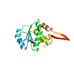 | | Crystal Structure of Cydia Pomonella PTP-2 phosphatase | | 分子名称: | CALCIUM ION, GLYCEROL, ORF98 PTP-2 | | 著者 | Huang, G, Keown, J.P, Oliver, M.R, Metcalf, P. | | 登録日 | 2018-10-31 | | 公開日 | 2019-02-20 | | 最終更新日 | 2024-05-15 | | 実験手法 | X-RAY DIFFRACTION (1.65 Å) | | 主引用文献 | Crystal structure of protein tyrosine phosphatase-2 from Cydia pomonella granulovirus.
Acta Crystallogr.,Sect.F, 75, 2019
|
|
1JPE
 | |
1JZD
 | | DsbC-DsbDalpha complex | | 分子名称: | thiol:disulfide interchange protein dsbc, thiol:disulfide interchange protein dsbd | | 著者 | Haebel, P.W, Goldstone, D, Katzen, F, Beckwith, J, Metcalf, P. | | 登録日 | 2001-09-15 | | 公開日 | 2003-03-08 | | 最終更新日 | 2023-08-16 | | 実験手法 | X-RAY DIFFRACTION (2.3 Å) | | 主引用文献 | The Disulfide Bond Isomerase DsbC is Activated by an
Immunoglobulin-fold Thiol Oxidoreductase: Crystal structure of the
DsbC-DsbDalpha complex.
Embo J., 21, 2002
|
|
1JZO
 | | DsbC C101S | | 分子名称: | THIOL:DISULFIDE INTERCHANGE PROTEIN DSBC | | 著者 | Haebel, P.W, Goldstone, D, Katzen, F, Beckwith, J, Metcalf, P. | | 登録日 | 2001-09-17 | | 公開日 | 2003-03-08 | | 最終更新日 | 2023-08-16 | | 実験手法 | X-RAY DIFFRACTION (1.92 Å) | | 主引用文献 | The Disulfide Bond Isomerase DsbC is Activated by an
Immunoglobulin-fold Thiol Oxidoreductase: Crystal Structure of the
DsbC-DsbDalpha complex.
Embo J., 21, 2002
|
|
4YN2
 | | THE ATOMIC STRUCTURE OF WISEANA SPP ENTOMOPOXVIRUS (WSEPV) FUSOLIN SPINDLES | | 分子名称: | 1,2-ETHANEDIOL, FUSOLIN, ZINC ION, ... | | 著者 | Chiu, E, Bunker, R.D, Metcalf, P. | | 登録日 | 2015-03-09 | | 公開日 | 2015-04-08 | | 最終更新日 | 2020-07-29 | | 実験手法 | X-RAY DIFFRACTION (2.02 Å) | | 主引用文献 | Structural basis for the enhancement of virulence by viral spindles and their in vivo crystallization.
Proc.Natl.Acad.Sci.USA, 112, 2015
|
|
4YE7
 | |
4YN1
 | | THE ATOMIC STRUCTURE OF ANOMALA CUPREA ENTOMOPOXVIRUS (ACEPV) FUSOLIN SPINDLES | | 分子名称: | 1,2-ETHANEDIOL, Fusolin, beta-D-mannopyranose-(1-4)-2-acetamido-2-deoxy-beta-D-glucopyranose-(1-4)-2-acetamido-2-deoxy-beta-D-glucopyranose | | 著者 | Chiu, E, Bunker, R.D, Metcalf, P. | | 登録日 | 2015-03-09 | | 公開日 | 2015-04-08 | | 最終更新日 | 2023-09-27 | | 実験手法 | X-RAY DIFFRACTION (1.9 Å) | | 主引用文献 | Structural basis for the enhancement of virulence by viral spindles and their in vivo crystallization.
Proc.Natl.Acad.Sci.USA, 112, 2015
|
|
2IY2
 | |
2IYJ
 | |
1RN1
 | | THREE-DIMENSIONAL STRUCTURE OF GLN 25-RIBONUCLEASE T1 AT 1.84 ANGSTROMS RESOLUTION: STRUCTURAL VARIATIONS AT THE BASE RECOGNITION AND CATALYTIC SITES | | 分子名称: | RIBONUCLEASE T1 ISOZYME, SULFATE ION | | 著者 | Arni, R.K, Pal, G.P, Ravichandran, K.G, Tulinsky, A, Walz Junior, F.G, Metcalf, P. | | 登録日 | 1991-11-22 | | 公開日 | 1994-01-31 | | 最終更新日 | 2019-08-14 | | 実験手法 | X-RAY DIFFRACTION (1.84 Å) | | 主引用文献 | Three-dimensional structure of Gln25-ribonuclease T1 at 1.84-A resolution: structural variations at the base recognition and catalytic sites.
Biochemistry, 31, 1992
|
|
2HYX
 | |
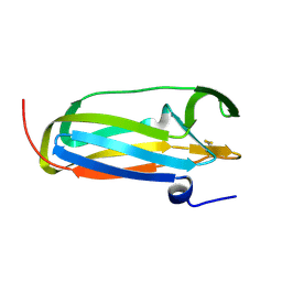
2VSG
 | | A Structural Motif in the Variant Surface Glycoproteins of Trypanosoma Brucei | | 分子名称: | VARIANT SURFACE GLYCOPROTEIN ILTAT 1.24 | | 著者 | Blum, M.L, Down, J.A, Metcalf, P, Freymann, D.M, Wiley, D.C. | | 登録日 | 1998-11-19 | | 公開日 | 1998-11-25 | | 最終更新日 | 2023-12-27 | | 実験手法 | X-RAY DIFFRACTION (2.7 Å) | | 主引用文献 | A structural motif in the variant surface glycoproteins of Trypanosoma brucei.
Nature, 362, 1993
|
|
2VOS
 | | Mycobacterium tuberculosis Folylpolyglutamate synthase complexed with ADP | | 分子名称: | ADENOSINE-5'-DIPHOSPHATE, COBALT (II) ION, FOLYLPOLYGLUTAMATE SYNTHASE PROTEIN FOLC, ... | | 著者 | Young, P.G, Baker, E.N, Metcalf, P, Smith, C.A. | | 登録日 | 2008-02-19 | | 公開日 | 2008-07-01 | | 最終更新日 | 2011-07-13 | | 実験手法 | X-RAY DIFFRACTION (2 Å) | | 主引用文献 | Structures of Mycobacterium Tuberculosisfolylpolyglutamate Synthase Complexed with Adp and Amppcp.
Acta Crystallogr.,Sect.D, 64, 2008
|
|


