1FWP
 
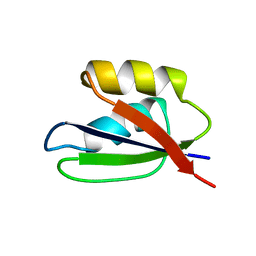 | |
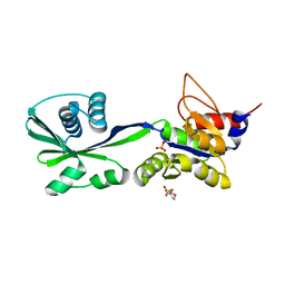
1EAY
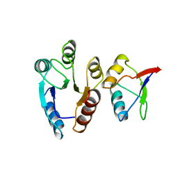 | | CHEY-BINDING (P2) DOMAIN OF CHEA IN COMPLEX WITH CHEY FROM ESCHERICHIA COLI | | 分子名称: | CHEA, CHEY | | 著者 | Mcevoy, M.M, Hausrath, A.C, Randolph, G.B, Remington, S.J, Dahlquist, F.W. | | 登録日 | 1998-04-23 | | 公開日 | 1998-07-15 | | 最終更新日 | 2024-05-22 | | 実験手法 | X-RAY DIFFRACTION (2 Å) | | 主引用文献 | Two binding modes reveal flexibility in kinase/response regulator interactions in the bacterial chemotaxis pathway.
Proc.Natl.Acad.Sci.USA, 95, 1998
|
|
6WJE
 | | Copper resistance protein copG- Form 2 | | 分子名称: | ACETATE ION, COPPER (II) ION, DUF411 domain-containing protein, ... | | 著者 | Hausrath, A.C, Ly, A.T, McEvoy, M.M. | | 登録日 | 2020-04-13 | | 公開日 | 2020-06-24 | | 最終更新日 | 2023-10-18 | | 実験手法 | X-RAY DIFFRACTION (2.5 Å) | | 主引用文献 | The bacterial copper resistance protein CopG contains a cysteine-bridged tetranuclear copper cluster.
J.Biol.Chem., 295, 2020
|
|
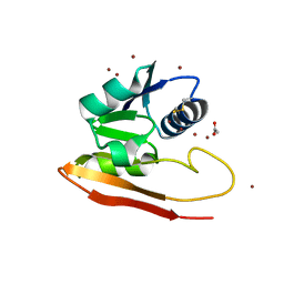
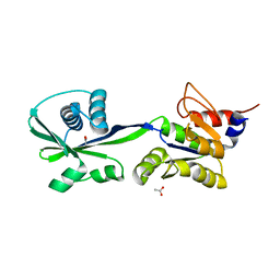
5KU5
 | |
1U8T
 | | Crystal structure of CheY D13K Y106W alone and in complex with a FliM peptide | | 分子名称: | Chemotaxis protein cheY, Flagellar motor switch protein fliM, SULFATE ION | | 著者 | Dyer, C.M, Quillin, M.L, Campos, A, Lu, J, McEvoy, M.M, Hausrath, A.C, Westbrook, E.M, Matsumura, P, Matthews, B.W, Dahlquist, F.W. | | 登録日 | 2004-08-06 | | 公開日 | 2004-10-05 | | 最終更新日 | 2021-10-20 | | 実験手法 | X-RAY DIFFRACTION (1.5 Å) | | 主引用文献 | Structure of the Constitutively Active Double Mutant CheY(D13K Y106W) Alone and in Complex with a FliM Peptide
J.Mol.Biol., 342, 2004
|
|
6WIS
 | | Copper resistance protein copG- Form 1 | | 分子名称: | ACETATE ION, COPPER (II) ION, DUF411 domain-containing protein, ... | | 著者 | Hausrath, A.C, Ly, A, McEvoy, M.M. | | 登録日 | 2020-04-10 | | 公開日 | 2020-06-24 | | 最終更新日 | 2020-08-19 | | 実験手法 | X-RAY DIFFRACTION (2 Å) | | 主引用文献 | The bacterial copper resistance protein CopG contains a cysteine-bridged tetranuclear copper cluster.
J.Biol.Chem., 295, 2020
|
|
1ZEQ
 | | 1.5 A Structure of apo-CusF residues 6-88 from Escherichia coli | | 分子名称: | Cation efflux system protein cusF | | 著者 | Loftin, I.R, Franke, S, Roberts, S.A, Weichsel, A, Heroux, A, Montfort, W.R, Rensing, C, McEvoy, M.M. | | 登録日 | 2005-04-19 | | 公開日 | 2005-08-02 | | 最終更新日 | 2024-02-14 | | 実験手法 | X-RAY DIFFRACTION (1.5 Å) | | 主引用文献 | A Novel Copper-Binding Fold for the Periplasmic Copper Resistance Protein CusF.
Biochemistry, 44, 2005
|
|
1C4W
 | | 1.9 A STRUCTURE OF A-THIOPHOSPHONATE MODIFIED CHEY D57C | | 分子名称: | CHEMOTAXIS PROTEIN CHEY | | 著者 | Halkides, C.J, McEvoy, M.M, Matsumura, P, Volz, K, Dahlquist, F.W. | | 登録日 | 1999-09-28 | | 公開日 | 2000-05-08 | | 最終更新日 | 2023-12-27 | | 実験手法 | X-RAY DIFFRACTION (1.84 Å) | | 主引用文献 | The 1.9 A resolution crystal structure of phosphono-CheY, an analogue of the active form of the response regulator, CheY.
Biochemistry, 39, 2000
|
|
3SKY
 | | 2.1A crystal structure of the phosphate bound ATP binding domain of Archaeoglobus fulgidus COPB | | 分子名称: | 3[N-MORPHOLINO]PROPANE SULFONIC ACID, Copper-exporting P-type ATPase B, PHOSPHATE ION | | 著者 | Jayakanthan, S, Roberts, S.A, Weichsel, A, Arguello, J.M, McEvoy, M.M. | | 登録日 | 2011-06-23 | | 公開日 | 2012-06-20 | | 最終更新日 | 2024-02-28 | | 実験手法 | X-RAY DIFFRACTION (2.1 Å) | | 主引用文献 | Conformations of the apo-, substrate-bound and phosphate-bound ATP-binding domain of the Cu(II) ATPase CopB illustrate coupling of domain movement to the catalytic cycle.
Biosci.Rep., 32, 2012
|
|
3SKX
 | | Crystal structure of the ATP binding domain of Archaeoglobus fulgidus COPB | | 分子名称: | ACETATE ION, Copper-exporting P-type ATPase B | | 著者 | Jayakanthan, S, Roberts, S.A, Weichsel, A, Arguello, J.M, McEvoy, M.M. | | 登録日 | 2011-06-23 | | 公開日 | 2012-06-20 | | 最終更新日 | 2024-02-28 | | 実験手法 | X-RAY DIFFRACTION (1.59 Å) | | 主引用文献 | Conformations of the apo-, substrate-bound and phosphate-bound ATP-binding domain of the Cu(II) ATPase CopB illustrate coupling of domain movement to the catalytic cycle.
Biosci.Rep., 32, 2012
|
|
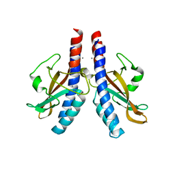
3E6Z
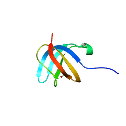 | |
2QCP
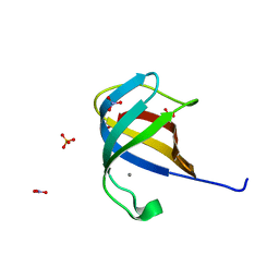 | |
1FQW
 | | CRYSTAL STRUCTURE OF ACTIVATED CHEY | | 分子名称: | BERYLLIUM TRIFLUORIDE ION, CHEMOTAXIS CHEY PROTEIN, MANGANESE (II) ION | | 著者 | Lee, S.Y, Cho, H.S, Pelton, J.G, Yan, D, Berry, E.A, Wemmer, D.E. | | 登録日 | 2000-09-07 | | 公開日 | 2001-07-18 | | 最終更新日 | 2024-02-07 | | 実験手法 | X-RAY DIFFRACTION (2.37 Å) | | 主引用文献 | Crystal structure of activated CheY. Comparison with other activated receiver domains.
J.Biol.Chem., 276, 2001
|
|


