6XJ7
 
 | |
6XJ6
 | |
8GLD
 | |
8GKD
 | |
4LMY
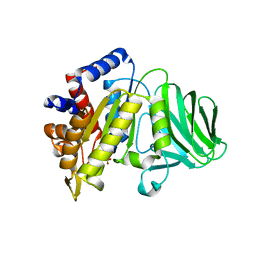
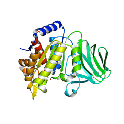
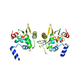
 | | Structure of GAS PerR-Zn-Zn | | 分子名称: | Peroxide stress regulator PerR, FUR family, ZINC ION | | 著者 | Lin, C.S, Chao, S.Y, Nix, J.C, Tseng, H.L, Tsou, C.C, Fei, C.H, Ciou, H.S, Jeng, U.S, Lin, Y.S, Chuang, W.J, Wu, J.J, Wang, S. | | 登録日 | 2013-07-11 | | 公開日 | 2014-04-02 | | 最終更新日 | 2024-03-20 | | 実験手法 | X-RAY DIFFRACTION (1.6 Å) | | 主引用文献 | Distinct structural features of the peroxide response regulator from group a streptococcus drive DNA binding
Plos One, 9, 2014
|
|
6DA1
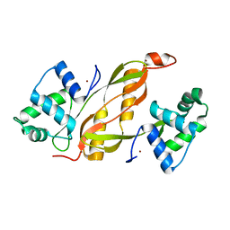
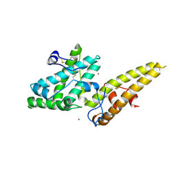
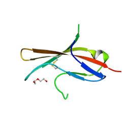
 | | ETS1 in complex with synthetic SRR mimic | | 分子名称: | Protein C-ets-1, SULFATE ION, serine-rich region (SRR) peptide | | 著者 | Perez-Borrajero, C, Okon, M, Lin, C.S, Scheu, K, Murphy, M.E.P, Graves, B.J, McIntosh, L.P. | | 登録日 | 2018-05-01 | | 公開日 | 2019-01-16 | | 最終更新日 | 2024-11-06 | | 実験手法 | X-RAY DIFFRACTION (2.000127 Å) | | 主引用文献 | The Biophysical Basis for Phosphorylation-Enhanced DNA-Binding Autoinhibition of the ETS1 Transcription Factor.
J. Mol. Biol., 431, 2019
|
|
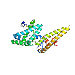
6DAT
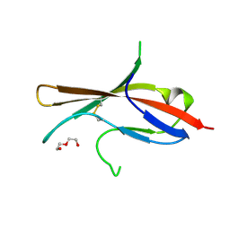
 | | ETS1 in complex with synthetic SRR mimic | | 分子名称: | Protein C-ets-1, SULFATE ION, serine-rich region (SRR) peptide | | 著者 | Perez-Borrajero, C, Okon, M, Lin, C.S, Scheu, K, Murphy, M.E.P, Graves, B.J, McIntosh, L.P. | | 登録日 | 2018-05-02 | | 公開日 | 2019-01-16 | | 最終更新日 | 2024-11-20 | | 実験手法 | X-RAY DIFFRACTION (2.35002637 Å) | | 主引用文献 | The Biophysical Basis for Phosphorylation-Enhanced DNA-Binding Autoinhibition of the ETS1 Transcription Factor.
J. Mol. Biol., 431, 2019
|
|
3VUV
 | | Crystal structure of the merozoite surface protein MSPDBL2 from P. falciparum bound to zinc | | 分子名称: | Erythrocyte membrane protein, putative, ZINC ION | | 著者 | Czabotar, P.E, Hodder, A.N, Clarke, O.B, Lin, C.S, Smith, B.J, Cowman, A.F. | | 登録日 | 2012-07-09 | | 公開日 | 2012-08-08 | | 最終更新日 | 2024-10-30 | | 実験手法 | X-RAY DIFFRACTION (2.114 Å) | | 主引用文献 | Insights into Duffy binding-like domains through the crystal structure and function of the merozoite surface protein MSPDBL2 from Plasmodium falciparum
J.Biol.Chem., 287, 2012
|
|
3VUU
 | | Crystal structure of the merozoite surface protein MSPDBL2 from P. falciparum | | 分子名称: | CHLORIDE ION, Erythrocyte membrane protein, putative | | 著者 | Czabotar, P.E, Hodder, A.N, Clarke, O.B, Lin, C.S, Smith, B.J, Cowman, A.F. | | 登録日 | 2012-07-09 | | 公開日 | 2012-08-08 | | 最終更新日 | 2024-11-13 | | 実験手法 | X-RAY DIFFRACTION (2.093 Å) | | 主引用文献 | Insights into Duffy binding-like domains through the crystal structure and function of the merozoite surface protein MSPDBL2 from Plasmodium falciparum
J.Biol.Chem., 287, 2012
|
|
5MJ0
 | |
5MJ1
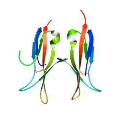
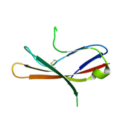
 | | Extracellular domain of human CD83 - rhombohedral crystal form | | 分子名称: | CD83 antigen, DI(HYDROXYETHYL)ETHER | | 著者 | Klingl, S, Egerer-Sieber, C, Schmid, B, Weiler, S, Muller, Y.A. | | 登録日 | 2016-11-29 | | 公開日 | 2017-03-29 | | 最終更新日 | 2024-11-20 | | 実験手法 | X-RAY DIFFRACTION (1.8 Å) | | 主引用文献 | Crystal Structure of the Extracellular Domain of the Human Dendritic Cell Surface Marker CD83.
J. Mol. Biol., 429, 2017
|
|
5MJ2
 | | Extracellular domain of human CD83 - rhombohedral crystal form after UV-RIP (S-SAD data) | | 分子名称: | CD83 antigen, DI(HYDROXYETHYL)ETHER | | 著者 | Klingl, S, Egerer-Sieber, C, Schmid, B, Weiler, S, Muller, Y.A. | | 登録日 | 2016-11-29 | | 公開日 | 2017-03-29 | | 最終更新日 | 2024-11-20 | | 実験手法 | X-RAY DIFFRACTION (1.98 Å) | | 主引用文献 | Crystal Structure of the Extracellular Domain of the Human Dendritic Cell Surface Marker CD83.
J. Mol. Biol., 429, 2017
|
|
5MIX
 | |
1FKD
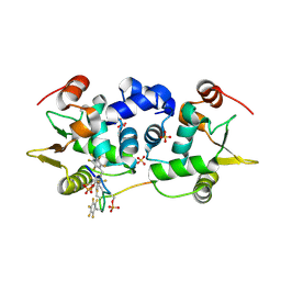
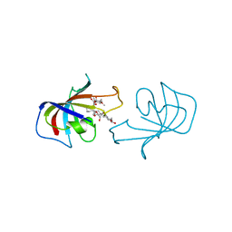 | | FK-506 BINDING PROTEIN: THREE-DIMENSIONAL STRUCTURE OF THE COMPLEX WITH THE ANTAGONIST L-685,818 | | 分子名称: | 18-HYDROXYASCOMYCIN, FK506 BINDING PROTEIN | | 著者 | Becker, J.W, Rotonda, J, Mckeever, B.M. | | 登録日 | 1992-12-02 | | 公開日 | 1994-01-31 | | 最終更新日 | 2024-02-07 | | 実験手法 | X-RAY DIFFRACTION (1.72 Å) | | 主引用文献 | FK-506-binding protein: three-dimensional structure of the complex with the antagonist L-685,818.
J.Biol.Chem., 268, 1993
|
|


