Loading
PDBj
メニューPDBj@FacebookPDBj@TwitterPDBj@YouTubewwPDB FoundationwwPDB
RCSB PDBPDBeBMRBAdv. SearchSearch help
Search by PDB author
2NBH
DownloadVisualize
BU of 2nbh by Molmil
Solution structure of the HYD1 hydrophobin from Schizophyllum commune
分子名称: Fungal hydrophobin
著者Langelaan, D, Smith, S, Grondin, J.
登録日2016-02-21
公開日2016-03-16
最終更新日2023-06-14
実験手法SOLUTION NMR
主引用文献Characterization of a Basidiomycota hydrophobin reveals the structural basis for a high-similarity Class I subdivision.
Sci Rep, 7, 2017
5IX9
DownloadVisualize
BU of 5ix9 by Molmil
Cell surface anchoring domain
分子名称: Antifreeze protein
著者Guo, S, Langelaan, D.
登録日2016-03-23
公開日2017-06-28
最終更新日2024-05-01
実験手法SOLUTION NMR
主引用文献Structure of a 1.5-MDa adhesin that binds its Antarctic bacterium to diatoms and ice.
Sci Adv, 3, 2017
2M8U
DownloadVisualize
BU of 2m8u by Molmil
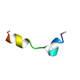
Solution structure of the Dictyostelium discodieum Myosin Light Chain, MlcC
分子名称: Myosin Light Chain, MlcC
著者Liburd, J.D, Miller, E, Langelaan, D, Chitayat, S, Crawley, S.W, Cote, G.P, Smith, S.P.
登録日2013-05-28
公開日2014-12-24
最終更新日2024-05-15
実験手法SOLUTION NMR
主引用文献Structure of the Single-lobe Myosin Light Chain C in Complex with the Light Chain-binding Domains of Myosin-1C Provides Insights into Divergent IQ Motif Recognition.
J.Biol.Chem., 291, 2016
2MNS
DownloadVisualize
BU of 2mns by Molmil
Solution NMR structure of the reovirus p15 fusion-associated small transmembrane (FAST) protein fusion-inducing lipid packing sensor (FLiPS) motif in dodecyl phosphocholine (DPC) micelles
分子名称: Membrane fusion protein p15
著者Sarker, M, Duncan, R, Read, J, Rainey, J.
登録日2014-04-09
公開日2014-04-23
実験手法SOLUTION NMR
主引用文献Novel helix-loop-helix fusion-inducing lipid packing sensor (FLiPS) for cell-cell fusion
To be Published

222415

件を2024-07-10に公開中

PDB statisticsPDBj update infoContact PDBjnumon