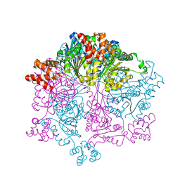
2P52
 
 | |
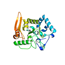
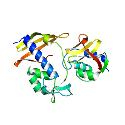
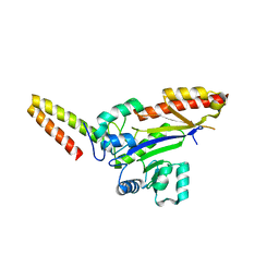
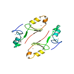
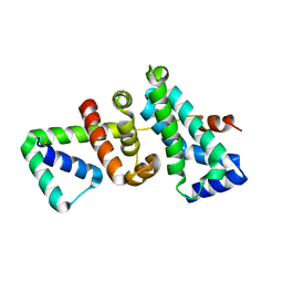
6JHE
 | | Crystal Structure of Bacillus subtilis SigW domain 4 in complexed with -35 element DNA | | 分子名称: | DNA (5'-D(*AP*AP*AP*GP*GP*TP*TP*TP*CP*AP*A)-3'), DNA (5'-D(P*TP*TP*GP*AP*AP*AP*CP*CP*TP*TP*T)-3'), ECF RNA polymerase sigma factor SigW | | 著者 | Kwon, E, Devkota, S.R, Pathak, D, Dahal, P, Kim, D.Y. | | 登録日 | 2019-02-18 | | 公開日 | 2020-01-01 | | 最終更新日 | 2023-11-22 | | 実験手法 | X-RAY DIFFRACTION (3.101 Å) | | 主引用文献 | Structural analysis of the recognition of the -35 promoter element by SigW from Bacillus subtilis.
Plos One, 14, 2019
|
|
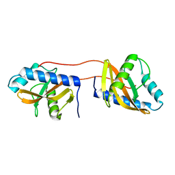
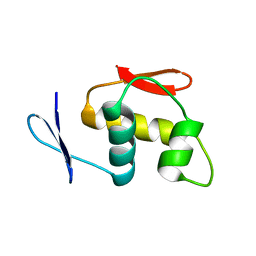
6JHK
 | |
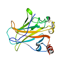
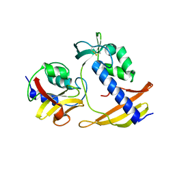
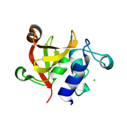
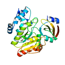
3OEO
 | | The crystal structure E. coli Spy | | 分子名称: | CADMIUM ION, Spheroplast protein Y | | 著者 | Kwon, E, Kim, D.Y, Gross, C.A, Gross, J.D, Kim, K.K. | | 登録日 | 2010-08-13 | | 公開日 | 2010-09-22 | | 最終更新日 | 2024-02-21 | | 実験手法 | X-RAY DIFFRACTION (2.7 Å) | | 主引用文献 | The crystal structure Escherichia coli Spy.
Protein Sci., 19, 2010
|
|
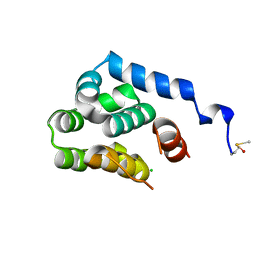
5X55
 | |
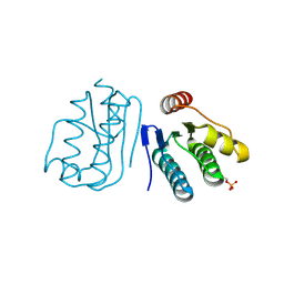
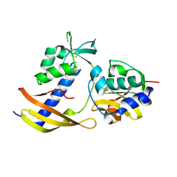
3V67
 | | Periplasmic domain of Vibrio parahaemolyticus CpxA | | 分子名称: | Sensor protein CpxA | | 著者 | Kwon, E, Kim, D.Y, Ngo, T.D, Gross, J.D, Kim, K.K. | | 登録日 | 2011-12-19 | | 公開日 | 2012-09-26 | | 最終更新日 | 2024-03-20 | | 実験手法 | X-RAY DIFFRACTION (2.3 Å) | | 主引用文献 | The crystal structure of the periplasmic domain of Vibrio parahaemolyticus CpxA
Protein Sci., 21, 2012
|
|
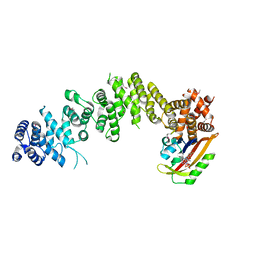
7W42
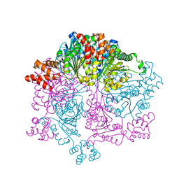 | | Crystal structure of Bacillus subtilis YjoB | | 分子名称: | Uncharacterized ATPase YjoB | | 著者 | Dahal, P, Kwon, E, Kim, D.Y. | | 登録日 | 2021-11-26 | | 公開日 | 2022-10-19 | | 最終更新日 | 2024-05-29 | | 実験手法 | X-RAY DIFFRACTION (2.619 Å) | | 主引用文献 | Crystal structure and biochemical analysis suggest that YjoB ATPase is a putative substrate-specific molecular chaperone.
Proc.Natl.Acad.Sci.USA, 119, 2022
|
|
7W43
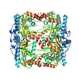 | |
7W46
 | |
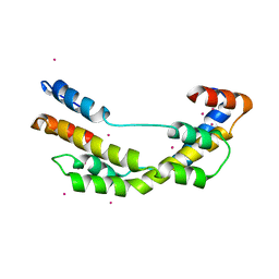
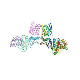
7WA4
 | | Crystal structure of GIGANTEA in complex with LKP2 | | 分子名称: | Adagio protein 2, FLAVIN MONONUCLEOTIDE, Protein GIGANTEA | | 著者 | Pathak, D, Dahal, P, Kwon, E, Kim, D.Y. | | 登録日 | 2021-12-12 | | 公開日 | 2022-04-27 | | 最終更新日 | 2022-05-04 | | 実験手法 | X-RAY DIFFRACTION (3.5 Å) | | 主引用文献 | Structural analysis of the regulation of blue-light receptors by GIGANTEA.
Cell Rep, 39, 2022
|
|
8I2E
 | |
8I2D
 | |
8I2F
 | |
6LYD
 | |
6M37
 | |
6M36
 | |
8WTC
 | |
8WT3
 | |
8WT4
 | |
8WTB
 | |
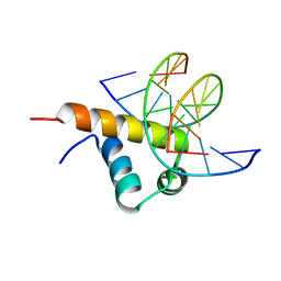
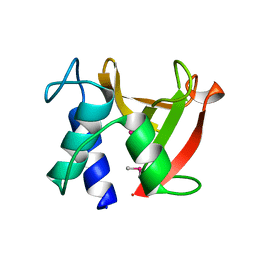
7WSJ
 | | Crystal structure of the tandem B-box domain of Arabidopsis thaliana CONSTANS | | 分子名称: | ZINC ION, Zinc finger protein CONSTANS | | 著者 | Dahal, P, Pathak, D, Kwon, E, Kim, D.Y. | | 登録日 | 2022-01-29 | | 公開日 | 2022-03-02 | | 最終更新日 | 2024-05-29 | | 実験手法 | X-RAY DIFFRACTION (2.4 Å) | | 主引用文献 | Crystal structure of a tandem B-box domain from Arabidopsis CONSTANS.
Biochem.Biophys.Res.Commun., 599, 2022
|
|
7CX5
 | |
2O7A
 | | T4 lysozyme C-terminal fragment | | 分子名称: | ACETATE ION, CHLORIDE ION, Lysozyme | | 著者 | Echols, N, Kwon, E, Marqusee, S.M, Alber, T. | | 登録日 | 2006-12-10 | | 公開日 | 2007-04-10 | | 最終更新日 | 2023-08-30 | | 実験手法 | X-RAY DIFFRACTION (0.84 Å) | | 主引用文献 | Exploring subdomain cooperativity in T4 lysozyme I: Structural and energetic studies of a circular permutant and protein fragment.
Protein Sci., 16, 2007
|
|
6LYE
 | |
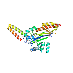
5WUR
 | | Crystal structure of SigW in complex with its anti-sigma RsiW, an oxdized form | | 分子名称: | Anti-sigma-W factor RsiW, ECF RNA polymerase sigma factor SigW | | 著者 | Devkota, S.R, Kwon, E, Ha, S.C, Kim, D.Y. | | 登録日 | 2016-12-20 | | 公開日 | 2017-03-29 | | 実験手法 | X-RAY DIFFRACTION (2.6 Å) | | 主引用文献 | Structural insights into the regulation of Bacillus subtilis SigW activity by anti-sigma RsiW
PLoS ONE, 12, 2017
|
|


