Loading
PDBj
メニューPDBj@FacebookPDBj@TwitterPDBj@YouTubewwPDB FoundationwwPDB
RCSB PDBPDBeBMRBAdv. SearchSearch help
Search by PDB author
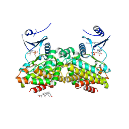
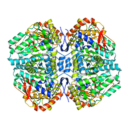
2C44
DownloadVisualize
BU of 2c44 by Molmil
Crystal Structure of E. coli Tryptophanase
分子名称: POTASSIUM ION, SULFATE ION, TRYPTOPHANASE
著者Ku, S.-Y, Yip, P, Howell, P.L.
登録日2005-10-15
公開日2006-06-28
最終更新日2023-12-13
実験手法X-RAY DIFFRACTION (2.8 Å)
主引用文献Structure of Escherichia Coli Tryptophanase
Acta Crystallogr.,Sect.D, 62, 2006
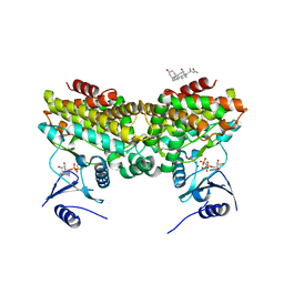
2PUL
DownloadVisualize
BU of 2pul by Molmil
Structures of 5-methylthioribose kinase reveal substrate specificity and unusual mode of nucleotide binding
分子名称: 3-[(3-CHOLAMIDOPROPYL)DIMETHYLAMMONIO]-1-PROPANESULFONATE, MAGNESIUM ION, Methylthioribose kinase, ...
著者Ku, S.-Y.
登録日2007-05-09
公開日2007-05-22
最終更新日2024-02-21
実験手法X-RAY DIFFRACTION (2 Å)
主引用文献Structures of 5-methylthioribose kinase reveal substrate specificity and unusual mode of nucleotide binding
J.Biol.Chem., 282, 2007
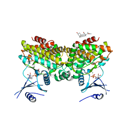
2PUP
DownloadVisualize
BU of 2pup by Molmil
Structures of 5-methylthioribose kinase reveal substrate specificity and unusual mode of nucleotide binding
分子名称: 3-[(3-CHOLAMIDOPROPYL)DIMETHYLAMMONIO]-1-PROPANESULFONATE, ADENOSINE-5'-DIPHOSPHATE, MAGNESIUM ION, ...
著者Ku, S.-Y.
登録日2007-05-09
公開日2007-05-22
最終更新日2024-02-21
実験手法X-RAY DIFFRACTION (2.6 Å)
主引用文献Structures of 5-methylthioribose kinase reveal substrate specificity and unusual mode of nucleotide binding
J.Biol.Chem., 282, 2007
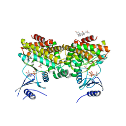
2PUN
DownloadVisualize
BU of 2pun by Molmil
Structures of 5-methylthioribose kinase reveal substrate specificity and unusual mode of nucleotide binding
分子名称: 3-[(3-CHOLAMIDOPROPYL)DIMETHYLAMMONIO]-1-PROPANESULFONATE, 5-S-methyl-5-thio-alpha-D-ribofuranose, MAGNESIUM ION, ...
著者Ku, S.-Y.
登録日2007-05-09
公開日2007-05-22
最終更新日2024-02-21
実験手法X-RAY DIFFRACTION (2.3 Å)
主引用文献Structures of 5-methylthioribose kinase reveal substrate specificity and unusual mode of nucleotide binding
J.Biol.Chem., 282, 2007
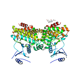
2PU8
DownloadVisualize
BU of 2pu8 by Molmil
Structures of 5-methylthioribose kinase reveal substrate specificity and unusual mode of nucleotide binding
分子名称: 3-[(3-CHOLAMIDOPROPYL)DIMETHYLAMMONIO]-1-PROPANESULFONATE, Methylthioribose kinase
著者Ku, S.-Y.
登録日2007-05-09
公開日2007-05-22
最終更新日2024-02-21
実験手法X-RAY DIFFRACTION (2.1 Å)
主引用文献Structures of 5-methylthioribose kinase reveal substrate specificity and unusual mode of nucleotide binding
J.Biol.Chem., 282, 2007
2PUI
DownloadVisualize
BU of 2pui by Molmil
Structures of 5-methylthioribose kinase reveal substrate specificity and unusual mode of nucleotide binding
分子名称: 3-[(3-CHOLAMIDOPROPYL)DIMETHYLAMMONIO]-1-PROPANESULFONATE, ADENOSINE-5'-DIPHOSPHATE, MAGNESIUM ION, ...
著者Ku, S.-Y.
登録日2007-05-09
公開日2007-05-22
最終更新日2024-02-21
実験手法X-RAY DIFFRACTION (2.2 Å)
主引用文献Structures of 5-methylthioribose kinase reveal substrate specificity and unusual mode of nucleotide binding
J.Biol.Chem., 282, 2007
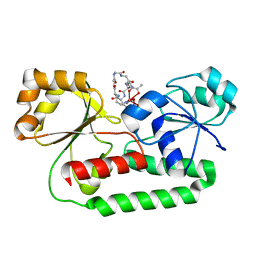
1EFD
DownloadVisualize
BU of 1efd by Molmil
PERIPLASMIC FERRIC SIDEROPHORE BINDING PROTEIN FHUD COMPLEXED WITH GALLICHROME
分子名称: FERRICHROME-BINDING PERIPLASMIC PROTEIN, GALLICHROME
著者Clarke, T.E, Ku, S.-Y, Dougan, D.R, Vogel, H.J, Tari, L.W.
登録日2000-02-07
公開日2000-04-05
最終更新日2024-02-07
実験手法X-RAY DIFFRACTION (1.9 Å)
主引用文献The structure of the ferric siderophore binding protein FhuD complexed with gallichrome.
Nat.Struct.Biol., 7, 2000
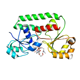
1ESZ
DownloadVisualize
BU of 1esz by Molmil
STRUCTURE OF THE PERIPLASMIC FERRIC SIDEROPHORE BINDING PROTEIN FHUD COMPLEXED WITH COPROGEN
分子名称: COPROGEN, FERRICHROME-BINDING PERIPLASMIC PROTEIN
著者Clarke, T.E, Braun, V, Winkelmann, G, Tari, L.W, Vogel, H.J.
登録日2000-04-11
公開日2002-04-17
最終更新日2024-02-07
実験手法X-RAY DIFFRACTION (2 Å)
主引用文献X-ray crystallographic structures of the Escherichia coli periplasmic protein FhuD bound to hydroxamate-type siderophores and the antibiotic albomycin.
J.Biol.Chem., 277, 2002
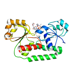
1K7S
DownloadVisualize
BU of 1k7s by Molmil
FhuD complexed with albomycin-delta 2
分子名称: DELTA-2-ALBOMYCIN A1, Ferrichrome-binding periplasmic protein
著者Clarke, T.E, Braun, V, Winkelmann, G, Tari, L.W, Vogel, H.J.
登録日2001-10-21
公開日2002-04-17
最終更新日2023-08-16
実験手法X-RAY DIFFRACTION (2.6 Å)
主引用文献X-ray crystallographic structures of the Escherichia coli periplasmic protein FhuD bound to hydroxamate-type siderophores and the antibiotic albomycin.
J.Biol.Chem., 277, 2002

222415

件を2024-07-10に公開中

PDB statisticsPDBj update infoContact PDBjnumon