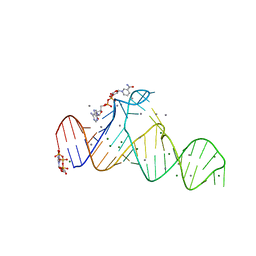
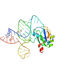
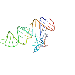
6UF1
 
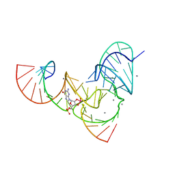 | | Pistol ribozyme transition-state analog vanadate | | 分子名称: | MANGANESE (II) ION, RNA (5'-R(*UP*CP*UP*GP*CP*UP*CP*UP*CP*(GVA)*UP*CP*CP*AP*A)-3'), RNA (50-MER) | | 著者 | Teplova, M, Falschlunger, C, Krasheninina, O, Patel, D.J, Micura, R. | | 登録日 | 2019-09-23 | | 公開日 | 2019-12-18 | | 最終更新日 | 2024-03-13 | | 実験手法 | X-RAY DIFFRACTION (3.1 Å) | | 主引用文献 | Crucial Roles of Two Hydrated Mg2+Ions in Reaction Catalysis of the Pistol Ribozyme.
Angew.Chem.Int.Ed.Engl., 59, 2020
|
|
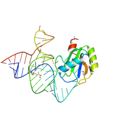
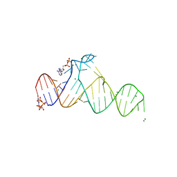
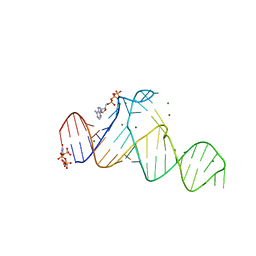
6UEY
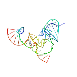 | | Pistol ribozyme transition-state analog vanadate | | 分子名称: | MAGNESIUM ION, RNA (5'-R(*UP*CP*UP*GP*CP*UP*CP*UP*CP*(GVA)*UP*CP*CP*AP*A)-3'), RNA (50-MER) | | 著者 | Teplova, M, Falschlunger, C, Krasheninina, O, Patel, D.J, Micura, R. | | 登録日 | 2019-09-23 | | 公開日 | 2019-12-18 | | 最終更新日 | 2024-03-13 | | 実験手法 | X-RAY DIFFRACTION (2.8 Å) | | 主引用文献 | Crucial Roles of Two Hydrated Mg2+Ions in Reaction Catalysis of the Pistol Ribozyme.
Angew.Chem.Int.Ed.Engl., 59, 2020
|
|
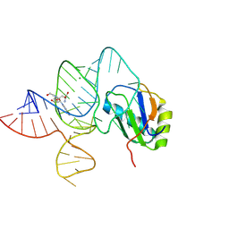
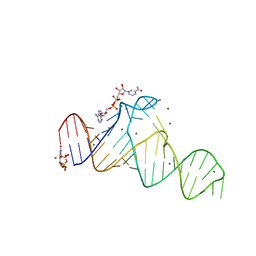
6UFJ
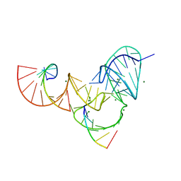 | | Pistol ribozyme product crystal structure | | 分子名称: | MAGNESIUM ION, RNA (5'-R(*UP*CP*CP*AP*G)-3'), RNA (5'-R(*UP*CP*UP*GP*CP*UP*CP*UP*CP*(23G))-3'), ... | | 著者 | Teplova, M, Falschlunger, C, Krasheninina, O, Patel, D.J, Micura, R. | | 登録日 | 2019-09-24 | | 公開日 | 2019-12-18 | | 最終更新日 | 2024-03-13 | | 実験手法 | X-RAY DIFFRACTION (2.645 Å) | | 主引用文献 | Crucial Roles of Two Hydrated Mg2+Ions in Reaction Catalysis of the Pistol Ribozyme.
Angew.Chem.Int.Ed.Engl., 59, 2020
|
|
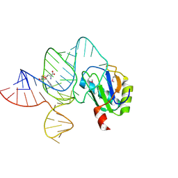
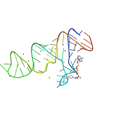
6UFK
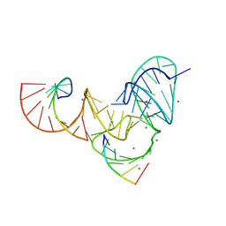 | | Pistol ribozyme product crystal soaked in Mn2+ | | 分子名称: | MANGANESE (II) ION, RNA (5'-R(*UP*CP*CP*AP*G)-3'), RNA (5'-R(*UP*CP*UP*GP*CP*UP*CP*UP*CP*(23G))-3'), ... | | 著者 | Teplova, M, Falschlunger, C, Krasheninina, O, Patel, D.J, Micura, R. | | 登録日 | 2019-09-24 | | 公開日 | 2019-12-18 | | 最終更新日 | 2024-03-13 | | 実験手法 | X-RAY DIFFRACTION (3.2 Å) | | 主引用文献 | Crucial Roles of Two Hydrated Mg2+Ions in Reaction Catalysis of the Pistol Ribozyme.
Angew.Chem.Int.Ed.Engl., 59, 2020
|
|
7D7V
 | |
7D7Z
 | |
6LAX
 | | the mutant SAM-VI riboswitch (U6C) bound to SAM | | 分子名称: | RNA (55-MER), S-ADENOSYLMETHIONINE, U1 small nuclear ribonucleoprotein A | | 著者 | Sun, A, Ren, A. | | 登録日 | 2019-11-13 | | 公開日 | 2020-01-01 | | 最終更新日 | 2023-11-22 | | 実験手法 | X-RAY DIFFRACTION (2.7 Å) | | 主引用文献 | SAM-VI riboswitch structure and signature for ligand discrimination.
Nat Commun, 10, 2019
|
|
6LAZ
 | | the wildtype SAM-VI riboswitch bound to a N-mustard SAM analog M1 | | 分子名称: | (2~{S})-4-[[(2~{R},3~{S},4~{R},5~{R})-5-(6-aminopurin-9-yl)-3,4-bis(oxidanyl)oxolan-2-yl]methyl-(2-hydroxyethyl)amino]-2-azaniumyl-butanoate, MAGNESIUM ION, RNA (55-MER), ... | | 著者 | Ren, A, Sun, A. | | 登録日 | 2019-11-13 | | 公開日 | 2020-01-01 | | 最終更新日 | 2023-11-22 | | 実験手法 | X-RAY DIFFRACTION (2.76 Å) | | 主引用文献 | SAM-VI riboswitch structure and signature for ligand discrimination.
Nat Commun, 10, 2019
|
|
6LAS
 | | the wildtype SAM-VI riboswitch bound to SAM | | 分子名称: | RNA (55-MER), S-ADENOSYLMETHIONINE, U1 small nuclear ribonucleoprotein A | | 著者 | Ren, A, Sun, A. | | 登録日 | 2019-11-13 | | 公開日 | 2020-01-01 | | 実験手法 | X-RAY DIFFRACTION (2.708 Å) | | 主引用文献 | SAM-VI riboswitch structure and signature for ligand discrimination.
Nat Commun, 10, 2019
|
|
6LAU
 | | the wildtype SAM-VI riboswitch bound to SAH | | 分子名称: | CESIUM ION, GUANOSINE-5'-TRIPHOSPHATE, RNA (54-MER), ... | | 著者 | Ren, A, Sun, A. | | 登録日 | 2019-11-13 | | 公開日 | 2020-01-01 | | 最終更新日 | 2023-11-22 | | 実験手法 | X-RAY DIFFRACTION (3.109 Å) | | 主引用文献 | SAM-VI riboswitch structure and signature for ligand discrimination.
Nat Commun, 10, 2019
|
|
7D7X
 | |
7D7W
 | |
7D82
 | |
7D81
 | |
7D7Y
 | |


