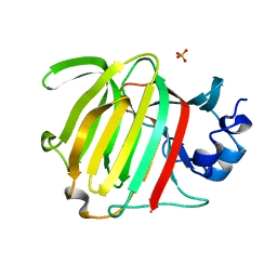
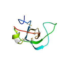
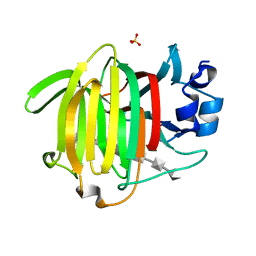
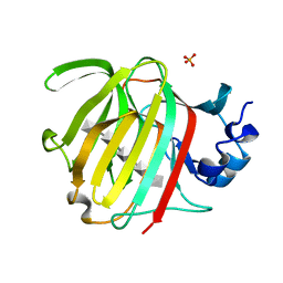
4UR4
 
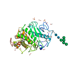
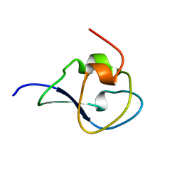 | | Structure of the type III fish antifreeze protein from Zoarces viviparus ZvAFP13 | | 分子名称: | ANTIFREEZE PROTEIN 13 | | 著者 | Wilkens, C, Poulsen, J.-C.N, Ramloev, H, Lo Leggio, L. | | 登録日 | 2014-06-26 | | 公開日 | 2014-07-23 | | 最終更新日 | 2024-01-10 | | 実験手法 | X-RAY DIFFRACTION (1.45 Å) | | 主引用文献 | Purification, Crystal Structure Determination and Functional Characterization of Type III Antifreeze Proteins from the European Eelpout Zoarces Viviparus.
Cryobiology, 69, 2014
|
|
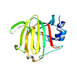
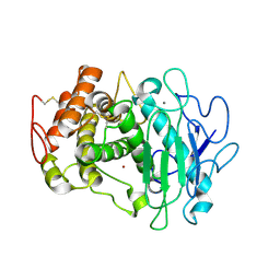
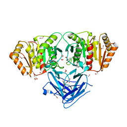
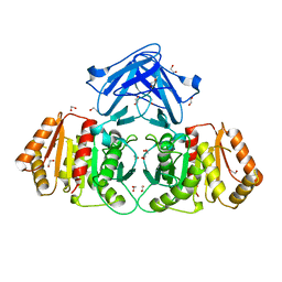
4UR6
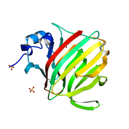
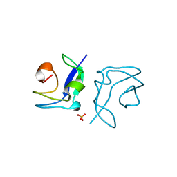 | | Structure of the type III fish antifreeze protein from Zoarces viviparus ZvAFP6 | | 分子名称: | SULFATE ION, TYPE III ANTIFREEZE PROTEIN 6 | | 著者 | Wilkens, C, Poulsen, J.-C.N, Ramloev, H, Lo Leggio, L. | | 登録日 | 2014-06-26 | | 公開日 | 2014-07-23 | | 最終更新日 | 2024-01-10 | | 実験手法 | X-RAY DIFFRACTION (1.2 Å) | | 主引用文献 | Purification, Crystal Structure Determination and Functional Characterization of Type III Antifreeze Proteins from the European Eelpout Zoarces Viviparus.
Cryobiology, 69, 2014
|
|
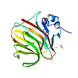
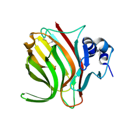
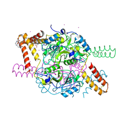
7PUG
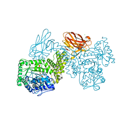 | | GH115 alpha-1,2-glucuronidase in complex with xylopentaose | | 分子名称: | CALCIUM ION, CHLORIDE ION, beta-D-xylopyranose-(1-4)-beta-D-xylopyranose-(1-4)-beta-D-xylopyranose-(1-4)-beta-D-xylopyranose-(1-4)-beta-D-xylopyranose, ... | | 著者 | Wilkens, C, Morth, J.P, Polikarpov, I. | | 登録日 | 2021-09-29 | | 公開日 | 2022-01-19 | | 最終更新日 | 2024-01-31 | | 実験手法 | X-RAY DIFFRACTION (2.66 Å) | | 主引用文献 | A GH115 alpha-glucuronidase structure reveals dimerization-mediated substrate binding and a proton wire potentially important for catalysis.
Acta Crystallogr D Struct Biol, 78, 2022
|
|
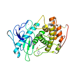
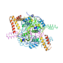
7PXQ
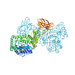 | | GH115 alpha-1,2-glucuronidase D303A | | 分子名称: | CALCIUM ION, xylan alpha-1,2-glucuronidase | | 著者 | Wilkens, C, Morth, J.P, Polikarpov, I. | | 登録日 | 2021-10-08 | | 公開日 | 2022-01-19 | | 最終更新日 | 2024-01-31 | | 実験手法 | X-RAY DIFFRACTION (2.3 Å) | | 主引用文献 | A GH115 alpha-glucuronidase structure reveals dimerization-mediated substrate binding and a proton wire potentially important for catalysis.
Acta Crystallogr D Struct Biol, 78, 2022
|
|
8P6O
 | |
8PED
 | |
8R43
 | |
8RBI
 | |
6E98
 | |
6E9M
 | |
7QFY
 | | Fusarium oxysporum M36 protease without the propeptide | | 分子名称: | 2-acetamido-2-deoxy-beta-D-glucopyranose-(1-4)-2-acetamido-2-deoxy-beta-D-glucopyranose, CALCIUM ION, Extracellular metalloproteinase, ... | | 著者 | Wilkens, C, Qiu, J, Meyer, A.S, Morth, J.P. | | 登録日 | 2021-12-07 | | 公開日 | 2022-12-21 | | 最終更新日 | 2024-01-31 | | 実験手法 | X-RAY DIFFRACTION (1.62 Å) | | 主引用文献 | Fusarium oxysporum M36 protease without the propeptide
To Be Published
|
|
6YWF
 | |
7QP3
 | | Pseudogymnoascus pannorum M36 protease without the propeptide | | 分子名称: | 2-acetamido-2-deoxy-alpha-D-galactopyranose, 4-(2-HYDROXYETHYL)-1-PIPERAZINE ETHANESULFONIC ACID, CALCIUM ION, ... | | 著者 | Wilkens, C, Qiu, J, Meyer, A.S, Morth, J.P. | | 登録日 | 2021-12-30 | | 公開日 | 2023-01-18 | | 最終更新日 | 2024-01-31 | | 実験手法 | X-RAY DIFFRACTION (1.85 Å) | | 主引用文献 | Phaeosphaeria nodorum M36 protease without the propeptide
To Be Published
|
|
8BBP
 | |
8BF3
 | |
8BJO
 | |
8BXZ
 | |
7Z6T
 | | Aspergillus clavatus M36 protease without the propeptide | | 分子名称: | 1,2-ETHANEDIOL, CALCIUM ION, Extracellular metalloproteinase mep, ... | | 著者 | Wilkens, C, Qiu, J, Meyer, A.S, Morth, J.P. | | 登録日 | 2022-03-14 | | 公開日 | 2023-03-22 | | 最終更新日 | 2024-02-07 | | 実験手法 | X-RAY DIFFRACTION (1.51 Å) | | 主引用文献 | Aspergillus clavatus M36 protease without the propeptide
To Be Published
|
|
8BZK
 | |
8C0M
 | |
1L8W
 | | Crystal Structure of Lyme Disease Variable Surface Antigen VlsE of Borrelia burgdorferi | | 分子名称: | VlsE1 | | 著者 | Eicken, C, Sharma, V, Klabunde, T, Lawrenz, M.B, Hardham, J.M, Norris, S.J, Sacchettini, J.C. | | 登録日 | 2002-03-21 | | 公開日 | 2002-06-19 | | 最終更新日 | 2018-03-07 | | 実験手法 | X-RAY DIFFRACTION (2.3 Å) | | 主引用文献 | Crystal structure of Lyme disease variable surface antigen VlsE of Borrelia burgdorferi.
J.Biol.Chem., 277, 2002
|
|
5NVD
 | | Crystal structure of hexameric CBS-CP12 protein from bloom-forming cyanobacteria at 2.5 A resolution in P6322 crystal form | | 分子名称: | CBS-CP12 | | 著者 | Hackenberg, C, Hakanpaa, J, Eigner, C, Antonyuk, S.V, Dittmann, E, Lamzin, V.S. | | 登録日 | 2017-05-04 | | 公開日 | 2018-05-30 | | 最終更新日 | 2024-01-17 | | 実験手法 | X-RAY DIFFRACTION (2.5 Å) | | 主引用文献 | Structural and functional insights into the unique CBS-CP12 fusion protein family in cyanobacteria.
Proc. Natl. Acad. Sci. U.S.A., 115, 2018
|
|
5NPL
 | | Crystal structure of hexameric CBS-CP12 protein from bloom-forming cyanobacteria, Yb-derivative at 2.8 A resolution | | 分子名称: | 10-((2R)-2-HYDROXYPROPYL)-1,4,7,10-TETRAAZACYCLODODECANE 1,4,7-TRIACETIC ACID, Similar to tr|Q8YYT1|Q8YYT1, YTTERBIUM (III) ION | | 著者 | Hackenberg, C, Hakanpaa, J, Antonyuk, S.V, Dittmann, E, Lamzin, V.S. | | 登録日 | 2017-04-17 | | 公開日 | 2018-05-30 | | 最終更新日 | 2018-07-11 | | 実験手法 | X-RAY DIFFRACTION (2.79 Å) | | 主引用文献 | Structural and functional insights into the unique CBS-CP12 fusion protein family in cyanobacteria.
Proc. Natl. Acad. Sci. U.S.A., 115, 2018
|
|
5NMU
 | | Structure of hexameric CBS-CP12 protein from bloom-forming cyanobacteria | | 分子名称: | CBS-CP12, CHLORIDE ION | | 著者 | Hackenberg, C, Hakanpaa, J, Antonyuk, S.V, Dittmann, E, Lamzin, V.S. | | 登録日 | 2017-04-07 | | 公開日 | 2018-05-16 | | 最終更新日 | 2024-01-17 | | 実験手法 | X-RAY DIFFRACTION (2.15 Å) | | 主引用文献 | Structural and functional insights into the unique CBS-CP12 fusion protein family in cyanobacteria.
Proc. Natl. Acad. Sci. U.S.A., 115, 2018
|
|
1R1V
 | | Crystal structure of the metal-sensing transcriptional repressor CzrA from Staphylococcus aureus in the Zn2-form | | 分子名称: | ZINC ION, repressor protein | | 著者 | Eicken, C, Pennella, M.A, Chen, X, Koshlap, K.M, VanZile, M.L, Sacchettini, J.C, Giedroc, D.P. | | 登録日 | 2003-09-25 | | 公開日 | 2004-05-18 | | 最終更新日 | 2023-08-23 | | 実験手法 | X-RAY DIFFRACTION (2.3 Å) | | 主引用文献 | A metal-ligand-mediated intersubunit allosteric switch in related SmtB/ArsR zinc sensor proteins.
J.Mol.Biol., 333, 2003
|
|


