Loading
PDBj
メニューPDBj@FacebookPDBj@TwitterPDBj@YouTubewwPDB FoundationwwPDB
RCSB PDBPDBeBMRBAdv. SearchSearch help
Search by PDB author
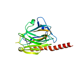
3POS
DownloadVisualize
BU of 3pos by Molmil
Crystal structure of the globular domain of human calreticulin
分子名称: CALCIUM ION, Calreticulin
著者Chouquet, A, Paidassi, H, Ling, W.-L, Frachet, P, Houen, G, Arlaud, G.J, Gaboriaud, C.
登録日2010-11-23
公開日2011-03-09
最終更新日2024-10-16
実験手法X-RAY DIFFRACTION (1.65 Å)
主引用文献X-ray structure of the human calreticulin globular domain reveals a Peptide-binding area and suggests a multi-molecular mechanism
Plos One, 6, 2011
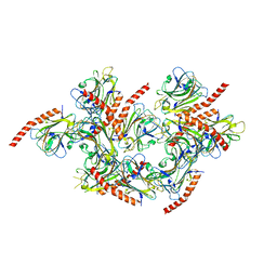
3POW
DownloadVisualize
BU of 3pow by Molmil
Crystal structure of the globular domain of human calreticulin
分子名称: CALCIUM ION, calreticulin
著者Gaboriaud, C.
登録日2010-11-23
公開日2011-03-09
最終更新日2024-10-30
実験手法X-RAY DIFFRACTION (1.55 Å)
主引用文献X-ray structure of the human calreticulin globular domain reveals a Peptide-binding area and suggests a multi-molecular mechanism
Plos One, 6, 2011
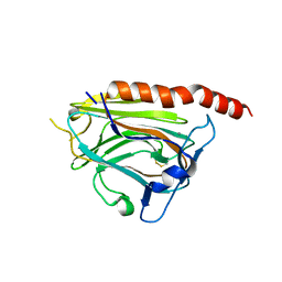
5LK5
DownloadVisualize
BU of 5lk5 by Molmil
Crystal structure of the globular domain of human calreticulin mutant D71K
分子名称: CALCIUM ION, CHLORIDE ION, Calreticulin,Calreticulin
著者Gaboriaud, C, Cioci, G.
登録日2016-07-21
公開日2016-08-31
最終更新日2019-10-16
実験手法X-RAY DIFFRACTION (2.3 Å)
主引用文献Structures of parasite calreticulins provide insights into their flexibility and dual carbohydrate/peptide-binding properties.
IUCrJ, 3, 2016

226707

件を2024-10-30に公開中

PDB statisticsPDBj update infoContact PDBjnumon