4N3D
 
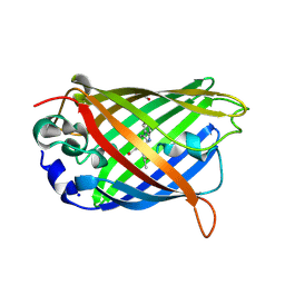
 | | Crystal structure of the dimeric variant EGFP-K162Q in P61 space group | | 分子名称: | GLYCEROL, Green fluorescent protein, PHOSPHATE ION, ... | | 著者 | Pletneva, N.V, Pletnev, V.Z, Pletnev, S.V. | | 登録日 | 2013-10-07 | | 公開日 | 2014-08-27 | | 実験手法 | X-RAY DIFFRACTION (1.34 Å) | | 主引用文献 | Three dimensional structure of the dimeric gene-engineered variant of green fluorescent protein egfp-K162Q in P61 crystal space group
Rus.J.Bioorg.Chem., 40, 2014
|
|
4RTC
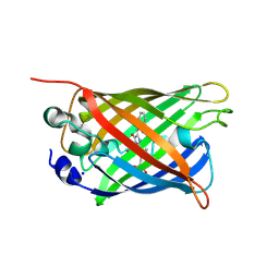
 | | Crystal structure of the green fluorescent variant, nowGFP, of the cyan Cerulean at pH 9.0 | | 分子名称: | GLYCEROL, nowGFP | | 著者 | Pletnev, V.Z, Pletneva, N.V, Pletnev, S.V. | | 登録日 | 2014-11-14 | | 公開日 | 2015-09-02 | | 最終更新日 | 2023-12-06 | | 実験手法 | X-RAY DIFFRACTION (1.35 Å) | | 主引用文献 | Structure of the green fluorescent protein NowGFP with an anionic tryptophan-based chromophore.
Acta Crystallogr.,Sect.D, 71, 2015
|
|
4RYS
 | |
4RYW
 | |
5DRF
 | |
5DQB
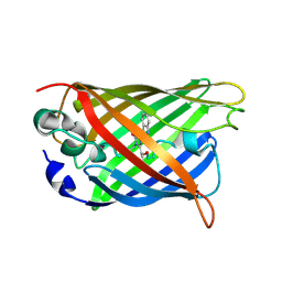
 | | Green/cyan WasCFP at pH 8.0 | | 分子名称: | GLYCEROL, SODIUM ION, WasCFP_pH2 | | 著者 | Pletnev, V, Pletneva, N, Pletnev, S. | | 登録日 | 2015-09-14 | | 公開日 | 2016-07-27 | | 最終更新日 | 2019-02-20 | | 実験手法 | X-RAY DIFFRACTION (1.25 Å) | | 主引用文献 | Crystal structure of pH and T dependent green fluorescent protein WasCFP with Trp based chromophore
Russ.J.Bioorganic Chem., 42 (6), 2016
|
|
5DQM
 | |
3LVD
 | |
3LVC
 | |
3LVA
 | |
5DRG
 | | Green/cyan WasCFP at pH 10.0 | | 分子名称: | GLYCEROL, Green/cyan WasCFP_pH10 at pH 10.0, SODIUM ION | | 著者 | Pletnev, V.Z, Pletneva, N.V, Pletnev, S.V. | | 登録日 | 2015-09-15 | | 公開日 | 2016-07-27 | | 最終更新日 | 2024-01-10 | | 実験手法 | X-RAY DIFFRACTION (1.14 Å) | | 主引用文献 | Crystal structure of pH and T dependent green fluorescent protein WasCFP with Trp based chromophore
Russ.J.Bioorganic Chem., 42 (6), 2016
|
|
1S5I
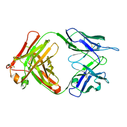 | | Fab (LNKB-2) of monoclonal antibody to Human Interleukin-2, crystal structure | | 分子名称: | Fab-fragment of monoclonal antibody | | 著者 | Pletnev, V.Z, Goryacheva, E.A, Tsygannik, I.N, Nesmeyanov, V.A, Pletnev, S.V, Pangborn, W, Duax, W. | | 登録日 | 2004-01-21 | | 公開日 | 2004-05-25 | | 最終更新日 | 2023-08-23 | | 実験手法 | X-RAY DIFFRACTION (2.7 Å) | | 主引用文献 | [A new crystal form of the Fab fragment of a monoclonal antibody to human interleukin-2: the three-dimensional structure at 2.7 A resolution].
Bioorg. Khim., 30
|
|


