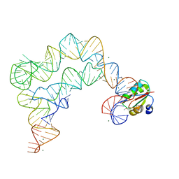
1AN4
 
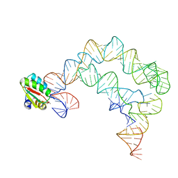
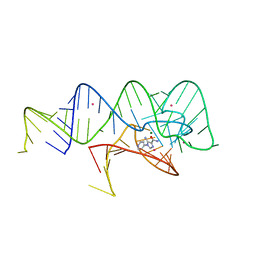
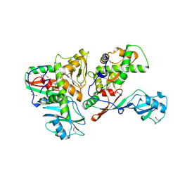
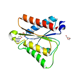
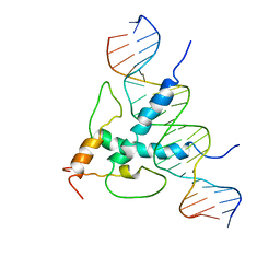 | | STRUCTURE AND FUNCTION OF THE B/HLH/Z DOMAIN OF USF | | 分子名称: | DNA (5'-D(*CP*AP*CP*CP*CP*GP*GP*TP*CP*AP*CP*GP*TP*GP*GP*CP*C P*TP*AP*CP*A)-3'), DNA (5'-D(*GP*TP*GP*TP*AP*GP*GP*CP*CP*AP*CP*GP*TP*GP*AP*CP*C P*GP*GP*GP*T)-3'), PROTEIN (UPSTREAM STIMULATORY FACTOR) | | 著者 | Ferre-D'Amare, A.R, Pognonec, P, Roeder, R.G, Burley, S.K. | | 登録日 | 1997-03-15 | | 公開日 | 1997-09-17 | | 最終更新日 | 2024-02-07 | | 実験手法 | X-RAY DIFFRACTION (2.9 Å) | | 主引用文献 | Structure and function of the b/HLH/Z domain of USF.
EMBO J., 13, 1994
|
|
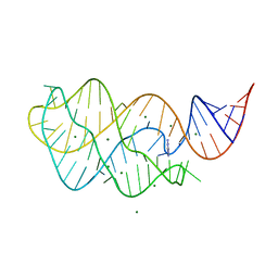
1DRZ
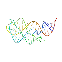
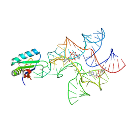
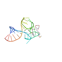
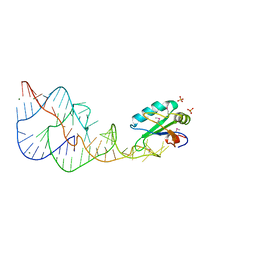 | | U1A SPLICEOSOMAL PROTEIN/HEPATITIS DELTA VIRUS GENOMIC RIBOZYME COMPLEX | | 分子名称: | MAGNESIUM ION, PROTEIN (U1 SMALL RIBONUCLEOPROTEIN A), RNA (HEPATITIS DELTA VIRUS GENOMIC RIBOZYME), ... | | 著者 | Ferre-D'Amare, A.R, Zhou, K, Doudna, J.A. | | 登録日 | 1998-09-01 | | 公開日 | 1999-02-16 | | 最終更新日 | 2021-11-03 | | 実験手法 | X-RAY DIFFRACTION (2.3 Å) | | 主引用文献 | Crystal structure of a hepatitis delta virus ribozyme.
Nature, 395, 1998
|
|
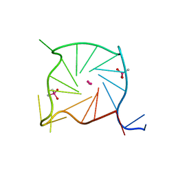
1AN2
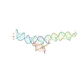
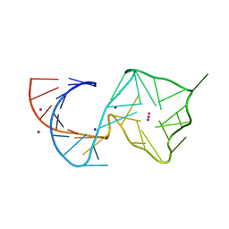
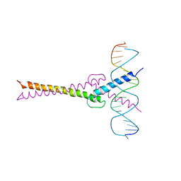 | | RECOGNITION BY MAX OF ITS COGNATE DNA THROUGH A DIMERIC B/HLH/Z DOMAIN | | 分子名称: | DNA (5'-D(*GP*TP*GP*TP*AP*GP*GP*TP*CP*AP*CP*GP*TP*GP*AP*CP*C P*TP*AP*CP*AP*C)- 3'), PROTEIN (TRANSCRIPTION FACTOR MAX (TF MAX)) | | 著者 | Ferre-D'Amare, A.R, Prendergast, G.C, Ziff, E.B, Burley, S.K. | | 登録日 | 1996-09-06 | | 公開日 | 1997-09-17 | | 最終更新日 | 2024-02-07 | | 実験手法 | X-RAY DIFFRACTION (2.9 Å) | | 主引用文献 | Recognition by Max of its cognate DNA through a dimeric b/HLH/Z domain.
Nature, 363, 1993
|
|
4TZP
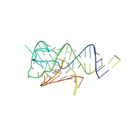
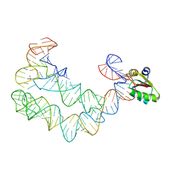 | | As Grown, Untreated Co-crystals of the Ternary Complex Containing a T-box Stem I RNA, its cognate tRNAGly, and B. subtilis YbxF protein | | 分子名称: | Ribosome-associated protein L7Ae-like, engineered tRNA, glyQ T-box Stem I | | 著者 | Zhang, J, Ferre-D'Amare, A.R. | | 登録日 | 2014-07-10 | | 公開日 | 2014-09-10 | | 最終更新日 | 2023-11-15 | | 実験手法 | X-RAY DIFFRACTION (8.503 Å) | | 主引用文献 | Dramatic Improvement of Crystals of Large RNAs by Cation Replacement and Dehydration.
Structure, 22, 2014
|
|
4TZV
 | | Co-crystals of the Ternary Complex Containing a T-box Stem I RNA, its Cognate tRNAGly, and B. subtilis YbxF protein, treated by removing lithium sulfate post crystallization | | 分子名称: | Ribosome-associated protein L7Ae-like, T-box stem I, engineered tRNA | | 著者 | Zhang, J, Ferre-D'Amare, A.R. | | 登録日 | 2014-07-11 | | 公開日 | 2014-09-10 | | 最終更新日 | 2023-11-15 | | 実験手法 | X-RAY DIFFRACTION (5.03 Å) | | 主引用文献 | Dramatic Improvement of Crystals of Large RNAs by Cation Replacement and Dehydration.
Structure, 22, 2014
|
|
4TZY
 | |
7MLW
 | |
6WZS
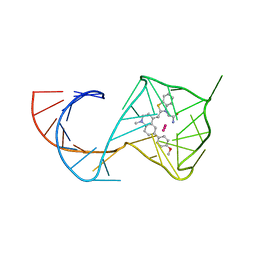
 | | Fusibacterium ulcerans ZTP riboswitch bound to m-1-pyridinyl AICA | | 分子名称: | 5-amino-1-(pyridin-3-yl)-1H-imidazole-4-carboxamide, Fusibacterium ulcerans ZTP riboswitch, MAGNESIUM ION, ... | | 著者 | Pichling, P, Jones, C.P, Ferre-D'Amare, A.R, Tran, B. | | 登録日 | 2020-05-14 | | 公開日 | 2021-03-24 | | 最終更新日 | 2023-10-18 | | 実験手法 | X-RAY DIFFRACTION (3.23 Å) | | 主引用文献 | Parallel Discovery Strategies Provide a Basis for Riboswitch Ligand Design.
Cell Chem Biol, 27, 2020
|
|
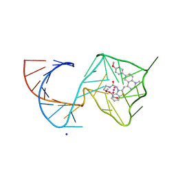
6WZR
 | | Fusibacterium ulcerans ZTP riboswitch bound to p-1-pyridinyl AICA | | 分子名称: | 5-amino-1-(pyridin-4-yl)-1H-imidazole-4-carboxamide, Fusibacterium ulcerans ZTP riboswitch, MAGNESIUM ION, ... | | 著者 | Pichling, P, Jones, C.P, Ferre-D'Amare, A.R, Tran, B. | | 登録日 | 2020-05-14 | | 公開日 | 2021-03-24 | | 最終更新日 | 2023-10-18 | | 実験手法 | X-RAY DIFFRACTION (3.2 Å) | | 主引用文献 | Parallel Discovery Strategies Provide a Basis for Riboswitch Ligand Design.
Cell Chem Biol, 27, 2020
|
|
4W90
 | | Crystal structure of Bacillus subtilis cyclic-di-AMP riboswitch ydaO | | 分子名称: | (2R,3R,3aS,5R,7aR,9R,10R,10aS,12R,14aR)-2,9-bis(6-amino-9H-purin-9-yl)octahydro-2H,7H-difuro[3,2-d:3',2'-j][1,3,7,9,2,8 ]tetraoxadiphosphacyclododecine-3,5,10,12-tetrol 5,12-dioxide, MAGNESIUM ION, U1 small nuclear ribonucleoprotein A, ... | | 著者 | Jones, C.P, Ferre-D'Amare, A.R. | | 登録日 | 2014-08-26 | | 公開日 | 2014-10-15 | | 最終更新日 | 2023-12-27 | | 実験手法 | X-RAY DIFFRACTION (3.118 Å) | | 主引用文献 | Crystal structure of a c-di-AMP riboswitch reveals an internally pseudo-dimeric RNA.
Embo J., 33, 2014
|
|
2AQA
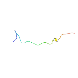 | |
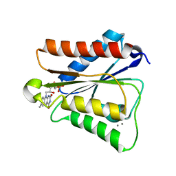
2AQC
 | | NMR Structural analysis of archaeal Nop10 | | 分子名称: | Ribosome biogenesis protein Nop10, ZINC ION | | 著者 | Hamma, T, Reichow, S.L, Varani, G, Ferre-D'Amare, A.R. | | 登録日 | 2005-08-17 | | 公開日 | 2005-11-15 | | 最終更新日 | 2024-05-22 | | 実験手法 | SOLUTION NMR | | 主引用文献 | The Cbf5-Nop10 complex is a molecular bracket that organizes box H/ACA RNPs.
Nat.Struct.Mol.Biol., 12, 2005
|
|
4W92
 | | Crystal structure of Bacillus subtilis cyclic-di-AMP riboswitch ydaO | | 分子名称: | (2R,3R,3aS,5R,7aR,9R,10R,10aS,12R,14aR)-2,9-bis(6-amino-9H-purin-9-yl)octahydro-2H,7H-difuro[3,2-d:3',2'-j][1,3,7,9,2,8 ]tetraoxadiphosphacyclododecine-3,5,10,12-tetrol 5,12-dioxide, 1,2-ETHANEDIOL, C-di-AMP ribsoswitch, ... | | 著者 | Jones, C.P, Ferre-D'Amare, A.R. | | 登録日 | 2014-08-26 | | 公開日 | 2014-10-22 | | 最終更新日 | 2023-12-27 | | 実験手法 | X-RAY DIFFRACTION (3.209 Å) | | 主引用文献 | Crystal structure of a c-di-AMP riboswitch reveals an internally pseudo-dimeric RNA.
Embo J., 33, 2014
|
|
4TZZ
 | | Co-crystals of the Ternary Complex Containing a T-box Stem I RNA, its Cognate tRNAGly, and B. subtilis YbxF protein, treated by removing lithium sulfate and increasing PEG3350 concentration from 20% to 45% post crystallization | | 分子名称: | MAGNESIUM ION, Ribosome-associated protein L7Ae-like, T-box Stem I RNA, ... | | 著者 | Zhang, J, Ferre-D'Amare, A.R. | | 登録日 | 2014-07-11 | | 公開日 | 2014-09-10 | | 最終更新日 | 2023-11-15 | | 実験手法 | X-RAY DIFFRACTION (3.64 Å) | | 主引用文献 | Dramatic Improvement of Crystals of Large RNAs by Cation Replacement and Dehydration.
Structure, 22, 2014
|
|
4TZX
 | |
7SXP
 | |
6PUP
 | |
1SB7
 | |
6PQ7
 | |
8U5P
 | | Structure of Mango II aptamer bound to T01-6A-B | | 分子名称: | 3-[2,16-dioxo-20-(2-oxohexahydro-1H-thieno[3,4-d]imidazol-4-yl)-6,9,12-trioxa-3,15-diazaicosan-1-yl]-2-{(E)-[6-(4-methoxyphenyl)-1-methylquinolin-4(1H)-ylidene]methyl}-1,3-benzothiazol-3-ium, Mango II, POTASSIUM ION | | 著者 | Passalacqua, L.F.M, Ferre-D'Amare, A.R. | | 登録日 | 2023-09-12 | | 公開日 | 2024-03-27 | | 実験手法 | X-RAY DIFFRACTION (2.9 Å) | | 主引用文献 | Structure of Mango II aptamer bound to T01-6A-B
To Be Published
|
|
8U5T
 | | Structure of Mango II variant aptamer bound to T01-6A-B | | 分子名称: | 3-{2,16-dioxo-20-[(3aS,4R,6aS)-2-oxohexahydro-1H-thieno[3,4-d]imidazol-4-yl]-6,9,12-trioxa-3,15-diazaicosan-1-yl}-2-{(E)-[6-(4-methoxyphenyl)-1-methylquinolin-4(1H)-ylidene]methyl}-1,3-benzothiazol-3-ium, Mango II variant, POTASSIUM ION, ... | | 著者 | Passalacqua, L.F.M, Ferre-D'Amare, A.R. | | 登録日 | 2023-09-12 | | 公開日 | 2024-03-27 | | 実験手法 | X-RAY DIFFRACTION (2.2 Å) | | 主引用文献 | Structure of Mango II variant aptamer bound to T01-6A-B
To Be Published
|
|
8U5Z
 | | Structure of Mango II variant aptamer bound to T01-7M-B | | 分子名称: | 2-[(E)-(1,7-dimethylquinolin-4(1H)-ylidene)methyl]-3-{2,16-dioxo-20-[(3aR,4S,6aR)-2-oxohexahydro-1H-thieno[3,4-d]imidazol-4-yl]-6,9,12-trioxa-3,15-diazaicosan-1-yl}-1,3-benzothiazol-3-ium, Mango II variant, POTASSIUM ION, ... | | 著者 | Passalacqua, L.F.M, Ferre-D'Amare, A.R. | | 登録日 | 2023-09-13 | | 公開日 | 2024-03-27 | | 実験手法 | X-RAY DIFFRACTION (2.5 Å) | | 主引用文献 | Structure of Mango II variant aptamer bound to T01-7M-B
To Be Published
|
|
6PUQ
 | |
8U5J
 | | Structure of Mango III variant aptamer bound to T01-07M-B | | 分子名称: | 2-[(E)-(1,7-dimethylquinolin-4(1H)-ylidene)methyl]-3-{2,16-dioxo-20-[(3aR,4S,6aR)-2-oxohexahydro-1H-thieno[3,4-d]imidazol-4-yl]-6,9,12-trioxa-3,15-diazaicosan-1-yl}-1,3-benzothiazol-3-ium, DIMETHYL SULFOXIDE, Mango III variant, ... | | 著者 | Passalacqua, L.F.M, Ferre-D'Amare, A.R. | | 登録日 | 2023-09-12 | | 公開日 | 2024-03-27 | | 実験手法 | X-RAY DIFFRACTION (1.7 Å) | | 主引用文献 | Structure of Mango III variant aptamer bound to T01-07M-B
To Be Published
|
|
8U5R
 | |


