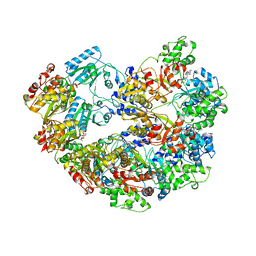
3M65
 
 | |
3M6A
 
 | |
3ZIH
 
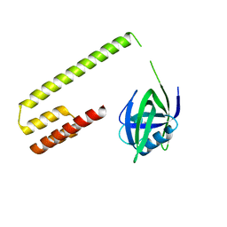
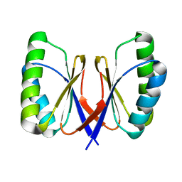
 | | Bacillus subtilis SepF, C-terminal domain | | 分子名称: | CELL DIVISION PROTEIN SEPF | | 著者 | Duman, R.E, Ishikawa, S, Celik, I, Ogasawara, N, Lowe, J, Hamoen, L.W. | | 登録日 | 2013-01-09 | | 公開日 | 2013-11-27 | | 最終更新日 | 2024-05-08 | | 実験手法 | X-RAY DIFFRACTION (2 Å) | | 主引用文献 | Structural and Genetic Analyses Reveal the Protein Sepf as a New Membrane Anchor for the Z Ring.
Proc.Natl.Acad.Sci.USA, 110, 2013
|
|
4CJ7
 
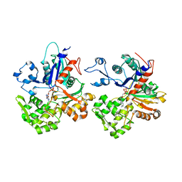
 | | Structure of Crenactin, an archeal actin-like protein | | 分子名称: | ACTIN/ACTIN FAMILY PROTEIN, ADENOSINE-5'-DIPHOSPHATE | | 著者 | Izore, T, Duman, R.E, Kureisaite-Ciziene, D, Lowe, J. | | 登録日 | 2013-12-19 | | 公開日 | 2014-01-22 | | 最終更新日 | 2023-12-20 | | 実験手法 | X-RAY DIFFRACTION (3.2 Å) | | 主引用文献 | Crenactin from Pyrobaculum Calidifontis is Closely Related to Actin in Structure and Forms Steep Helical Filaments.
FEBS Lett., 588, 2014
|
|
