7YSI
 
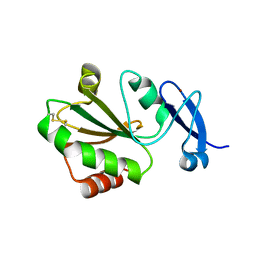 | | Crystal structure of thioredoxin 2 | | 分子名称: | Thiol disulfide reductase thioredoxin, ZINC ION | | 著者 | Chang, Y.J, Park, H.H. | | 登録日 | 2022-08-12 | | 公開日 | 2023-03-15 | | 最終更新日 | 2023-11-29 | | 実験手法 | X-RAY DIFFRACTION (1.202 Å) | | 主引用文献 | Comparison of the structure and activity of thioredoxin 2 and thioredoxin 1 from Acinetobacter baumannii.
Iucrj, 10, 2023
|
|
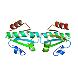
7XAO
 | |
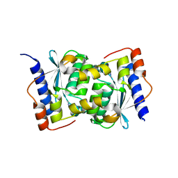
1WKQ
 | |
2GQG
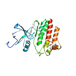 | | X-ray Crystal Structure of Dasatinib (BMS-354825) Bound to Activated ABL Kinase Domain | | 分子名称: | GLYCEROL, N-(2-CHLORO-6-METHYLPHENYL)-2-({6-[4-(2-HYDROXYETHYL)PIPERAZIN-1-YL]-2-METHYLPYRIMIDIN-4-YL}AMINO)-1,3-THIAZOLE-5-CARBOXAMIDE, Proto-oncogene tyrosine-protein kinase ABL1 | | 著者 | Klei, H.E. | | 登録日 | 2006-04-20 | | 公開日 | 2006-11-21 | | 最終更新日 | 2017-10-18 | | 実験手法 | X-RAY DIFFRACTION (2.4 Å) | | 主引用文献 | The Structure of Dasatinib (BMS-354825) Bound to Activated ABL Kinase Domain Elucidates Its Inhibitory Activity against Imatinib-Resistant ABL Mutants
CANCER RES., 66, 2006
|
|


