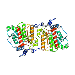
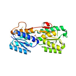
4XXH
 
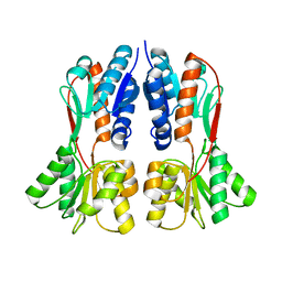 | | TREHALOSE REPRESSOR FROM ESCHERICHIA COLI | | 分子名称: | 6-O-phosphono-alpha-D-glucopyranose-(1-1)-alpha-D-glucopyranose, HTH-type transcriptional regulator TreR | | 著者 | Hars, U, Horlacher, R, Boos, W, Smart, O.S, Bricogne, G, Welte, W, Diederichs, K. | | 登録日 | 2015-01-30 | | 公開日 | 2015-02-11 | | 最終更新日 | 2024-05-08 | | 実験手法 | X-RAY DIFFRACTION (2.4 Å) | | 主引用文献 | CRYSTAL STRUCTURE OF THE EFFECTOR-BINDING DOMAIN OF THE TREHALOSE-REPRESSOR OF ESCHERICHIA COLI, A MEMBER OF THE LACI FAMILY, IN ITS COMPLEXES WITH INDUCER TREHALOSE-6-PHOSPHATE AND NONINDUCER TREHALOSE.
PROTEIN SCI., 7, 1998
|
|
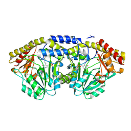
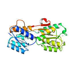
2F5T
 | | Crystal Structure of the sugar binding domain of the archaeal transcriptional regulator TrmB | | 分子名称: | IMIDAZOLE, alpha-D-glucopyranose-(1-4)-alpha-D-glucopyranose, archaeal transcriptional regulator TrmB | | 著者 | Krug, M, Lee, S.J, Diederichs, K, Boos, W, Welte, W. | | 登録日 | 2005-11-27 | | 公開日 | 2006-02-21 | | 最終更新日 | 2024-02-14 | | 実験手法 | X-RAY DIFFRACTION (1.45 Å) | | 主引用文献 | Crystal Structure of the Sugar Binding Domain of the Archaeal Transcriptional Regulator TrmB
J.Biol.Chem., 281, 2006
|
|
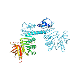
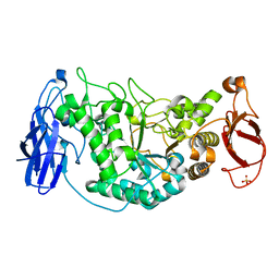
1Z6R
 | | Crystal structure of Mlc from Escherichia coli | | 分子名称: | Mlc protein, ZINC ION | | 著者 | Schiefner, A, Gerber, K, Seitz, S, Welte, W, Diederichs, K, Boos, W. | | 登録日 | 2005-03-23 | | 公開日 | 2005-06-14 | | 最終更新日 | 2021-11-10 | | 実験手法 | X-RAY DIFFRACTION (2.7 Å) | | 主引用文献 | The crystal structure of Mlc, a global regulator of sugar metabolism in Escherichia coli
J.Biol.Chem., 280, 2005
|
|
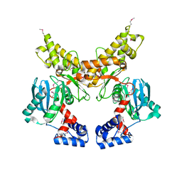
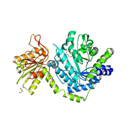
3QPH
 | | The three-dimensional structure of TrmB, a global transcriptional regulator of the hyperthermophilic archaeon Pyrococcus furiosus in complex with sucrose | | 分子名称: | ACETATE ION, GLYCEROL, TrmB, ... | | 著者 | Krug, M, Lee, S.-J, Boos, W, Welte, W, Diederichs, K. | | 登録日 | 2011-02-13 | | 公開日 | 2012-05-30 | | 最終更新日 | 2024-02-21 | | 実験手法 | X-RAY DIFFRACTION (2.992 Å) | | 主引用文献 | The three-dimensional structure of TrmB, a transcriptional regulator of dual function in the hyperthermophilic archaeon Pyrococcus furiosus in complex with sucrose.
Protein Sci., 22, 2013
|
|
2K14
 | | Solution structure of the soluble domain of the NfeD protein YuaF from Bacillus subtilis | | 分子名称: | YuaF protein | | 著者 | Walker, C.A, Hinderhofer, M, Witte, D.J, Boos, W, Moller, H.M. | | 登録日 | 2008-02-20 | | 公開日 | 2008-08-26 | | 最終更新日 | 2024-05-29 | | 実験手法 | SOLUTION NMR | | 主引用文献 | Solution structure of the soluble domain of the NfeD protein YuaF from Bacillus subtilis.
J.Biomol.Nmr, 42, 2008
|
|
1EU8
 | | STRUCTURE OF TREHALOSE MALTOSE BINDING PROTEIN FROM THERMOCOCCUS LITORALIS | | 分子名称: | CHLORIDE ION, PLATINUM (II) ION, TREHALOSE/MALTOSE BINDING PROTEIN, ... | | 著者 | Diez, J, Diederichs, K, Greller, G, Horlacher, R, Boos, W, Welte, W. | | 登録日 | 2000-04-14 | | 公開日 | 2001-03-14 | | 最終更新日 | 2024-02-07 | | 実験手法 | X-RAY DIFFRACTION (1.9 Å) | | 主引用文献 | The crystal structure of a liganded trehalose/maltose-binding protein from the hyperthermophilic Archaeon Thermococcus litoralis at 1.85 A.
J.Mol.Biol., 305, 2001
|
|
1G29
 | | MALK | | 分子名称: | 1,4-DIETHYLENE DIOXIDE, AMMONIUM ION, CHLORIDE ION, ... | | 著者 | Diederichs, K, Diez, J, Greller, G, Mueller, C, Breed, J, Schnell, C, Vonrhein, C, Boos, W, Welte, W. | | 登録日 | 2000-10-18 | | 公開日 | 2000-12-06 | | 最終更新日 | 2024-02-07 | | 実験手法 | X-RAY DIFFRACTION (1.9 Å) | | 主引用文献 | Crystal structure of MalK, the ATPase subunit of the trehalose/maltose ABC transporter of the archaeon Thermococcus litoralis.
EMBO J., 19, 2000
|
|
4KRX
 | | Structure of Aes from E. coli | | 分子名称: | Acetyl esterase, TETRAETHYLENE GLYCOL | | 著者 | Schiefner, A, Gerber, K, Brosig, A, Boos, W. | | 登録日 | 2013-05-17 | | 公開日 | 2013-08-21 | | 最終更新日 | 2023-09-20 | | 実験手法 | X-RAY DIFFRACTION (1.8 Å) | | 主引用文献 | Structural and mutational analyses of Aes, an inhibitor of MalT in Escherichia coli.
Proteins, 82, 2014
|
|
4KRY
 | | Structure of Aes from E. coli in covalent complex with PMS | | 分子名称: | Acetyl esterase, IMIDAZOLE, PENTAETHYLENE GLYCOL, ... | | 著者 | Schiefner, A, Gerber, K, Brosig, A, Boos, W. | | 登録日 | 2013-05-17 | | 公開日 | 2013-08-21 | | 最終更新日 | 2023-09-20 | | 実験手法 | X-RAY DIFFRACTION (2.3 Å) | | 主引用文献 | Structural and mutational analyses of Aes, an inhibitor of MalT in Escherichia coli.
Proteins, 82, 2014
|
|
1D2F
 | |
2WSK
 | | Crystal structure of Glycogen Debranching Enzyme GlgX from Escherichia coli K-12 | | 分子名称: | GLYCOGEN DEBRANCHING ENZYME, SULFATE ION | | 著者 | Song, H.-N, Park, J.-T, Jung, T.-Y, Park, K.-H, Woo, E.-J. | | 登録日 | 2009-09-08 | | 公開日 | 2010-09-01 | | 最終更新日 | 2023-12-20 | | 実験手法 | X-RAY DIFFRACTION (2.25 Å) | | 主引用文献 | Structural Rationale for the Short Branched Substrate Specificity of the Glycogen Debranching Enzyme Glgx.
Proteins, 78, 2010
|
|
3LK6
 | |
5BQT
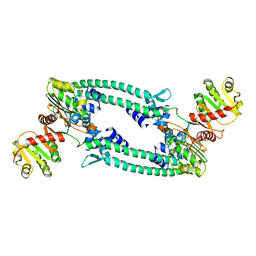 | | Structure of TrmBL2, an archaeal chromatin protein, shows a novel mode of DNA binding. | | 分子名称: | CALCIUM ION, Putative HTH-type transcriptional regulator TrmBL2 | | 著者 | Ahmad, M.U, Diederichs, K, Welte, W. | | 登録日 | 2015-05-29 | | 公開日 | 2015-09-02 | | 最終更新日 | 2024-01-10 | | 実験手法 | X-RAY DIFFRACTION (3 Å) | | 主引用文献 | Structural Insights into Nonspecific Binding of DNA by TrmBL2, an Archaeal Chromatin Protein.
J.Mol.Biol., 427, 2015
|
|
5BOX
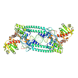 | | Structure of TrmBL2, an archaeal chromatin protein, shows a novel mode of DNA binding. | | 分子名称: | (4S)-2-METHYL-2,4-PENTANEDIOL, DNA (25-MER), DNA TGM (25-MER), ... | | 著者 | Ahmad, M.U, Diederichs, K, Welte, W. | | 登録日 | 2015-05-27 | | 公開日 | 2015-09-02 | | 最終更新日 | 2024-01-10 | | 実験手法 | X-RAY DIFFRACTION (2.5 Å) | | 主引用文献 | Structural Insights into Nonspecific Binding of DNA by TrmBL2, an Archaeal Chromatin Protein.
J.Mol.Biol., 427, 2015
|
|
5BPI
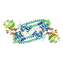 | | Structure of TrmBL2, an archaeal chromatin protein, shows a novel mode of DNA binding. | | 分子名称: | DNA (5'-D(P*TP*AP*TP*AP*TP*CP*AP*CP*TP*AP*TP*CP*GP*AP*TP*GP*AP*TP*AP*TP*A)-3'), DNA (5'-D(P*TP*AP*TP*AP*TP*CP*AP*TP*CP*GP*AP*TP*AP*GP*TP*GP*AP*TP*AP*TP*A)-3'), GLYCEROL, ... | | 著者 | Ahmad, M.U, Diederichs, K, Welte, W. | | 登録日 | 2015-05-28 | | 公開日 | 2015-09-02 | | 最終更新日 | 2024-01-10 | | 実験手法 | X-RAY DIFFRACTION (3.198 Å) | | 主引用文献 | Structural Insights into Nonspecific Binding of DNA by TrmBL2, an Archaeal Chromatin Protein.
J.Mol.Biol., 427, 2015
|
|
5BPD
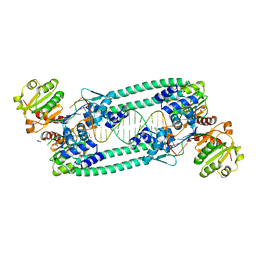 | | Structure of TrmBL2, an archaeal chromatin protein, shows a novel mode of DNA binding. | | 分子名称: | DNA (5'-D(P*TP*AP*TP*AP*TP*CP*AP*CP*TP*AP*TP*CP*GP*AP*TP*GP*AP*TP*AP*TP*A)-3'), DNA (5'-D(P*TP*AP*TP*AP*TP*CP*AP*TP*CP*GP*AP*TP*AP*GP*TP*GP*AP*TP*AP*TP*A)-3'), GLYCEROL, ... | | 著者 | Ahmad, M.U, Diederichs, K, Welte, W. | | 登録日 | 2015-05-28 | | 公開日 | 2015-09-02 | | 最終更新日 | 2024-05-08 | | 実験手法 | X-RAY DIFFRACTION (2.4 Å) | | 主引用文献 | Structural Insights into Nonspecific Binding of DNA by TrmBL2, an Archaeal Chromatin Protein.
J.Mol.Biol., 427, 2015
|
|
3GA5
 | | X-ray structure of glucose/galactose receptor from Salmonella typhimurium in complex with (2R)-glyceryl-beta-D-galactopyranoside | | 分子名称: | (2R)-2,3-dihydroxypropyl beta-D-galactopyranoside, CALCIUM ION, D-galactose-binding periplasmic protein, ... | | 著者 | Sooriyaarachchi, S, Ubhayasekera, W, Mowbray, S.L. | | 登録日 | 2009-02-16 | | 公開日 | 2009-04-14 | | 最終更新日 | 2023-11-01 | | 実験手法 | X-RAY DIFFRACTION (1.87 Å) | | 主引用文献 | X-ray structure of glucose/galactose receptor from Salmonella typhimurium in complex with the physiological ligand, (2R)-glyceryl-beta-D-galactopyranoside
Febs J., 276, 2009
|
|
3DZM
 | |
3GBP
 | |


