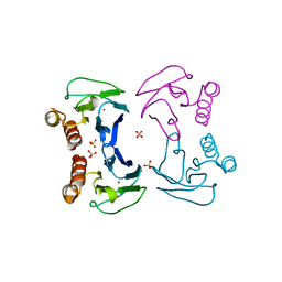
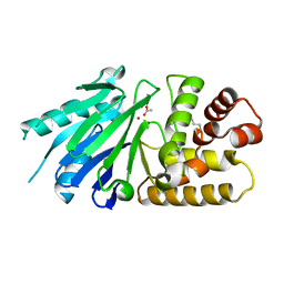
2WFB
 
 | | High resolution structure of the apo form of the orange protein (ORP) from Desulfovibrio gigas | | 分子名称: | ACETATE ION, PHOSPHATE ION, PUTATIVE UNCHARACTERIZED PROTEIN ORP | | 著者 | Najmudin, S, Bonifacio, C, Duarte, A.G, Pereira, A.S, Moura, I, Moura, J.M, Romao, M.J. | | 登録日 | 2009-04-03 | | 公開日 | 2010-05-26 | | 最終更新日 | 2023-12-13 | | 実験手法 | X-RAY DIFFRACTION (2 Å) | | 主引用文献 | High Resolution Crystal Structure of the Apo Form of the Orange Protein (Apo-Orp) from Desulfovibrio Gigas
To be Published
|
|
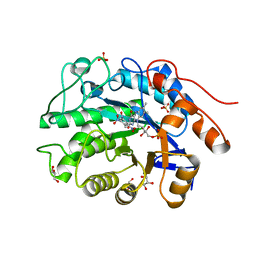
1Y07
 | | Crystal structure of the superoxide reductase from Treponema pallidum | | 分子名称: | FE (III) ION, MAGNESIUM ION, desulfoferrodoxin (rbo) | | 著者 | Santos-Silva, T, Trincao, J, Carvalho, A.L, Bonifacio, C, Auchere, F, Moura, J, Romao, M.J. | | 登録日 | 2004-11-15 | | 公開日 | 2005-11-22 | | 最終更新日 | 2024-03-13 | | 実験手法 | X-RAY DIFFRACTION (1.55 Å) | | 主引用文献 | The first crystal structure of class III superoxide reductase from Treponema pallidum.
J.Biol.Inorg.Chem., 11, 2006
|
|
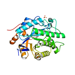
1RZ5
 | | Di-haem Cytochrome c Peroxidase, Form OUT | | 分子名称: | CALCIUM ION, Cytochrome c peroxidase, HEME C | | 著者 | Dias, J.M, Alves, T, Bonifacio, C, Pereira, A.S, Bourgeois, D, Moura, I, Romao, M.J. | | 登録日 | 2003-12-24 | | 公開日 | 2004-06-29 | | 最終更新日 | 2023-08-23 | | 実験手法 | X-RAY DIFFRACTION (2.4 Å) | | 主引用文献 | Structural basis for the mechanism of Ca(2+) activation of the di-heme cytochrome c peroxidase from Pseudomonas nautica 617.
Structure, 12, 2004
|
|
1RZ6
 | | Di-haem Cytochrome c Peroxidase, Form IN | | 分子名称: | CITRIC ACID, Cytochrome c peroxidase, HEME C | | 著者 | Dias, J.M, Alves, T, Bonifacio, C, Pereira, A, Bourgeois, D, Moura, I, Romao, M.J. | | 登録日 | 2003-12-24 | | 公開日 | 2004-06-29 | | 最終更新日 | 2023-08-23 | | 実験手法 | X-RAY DIFFRACTION (2.2 Å) | | 主引用文献 | Structural basis for the mechanism of Ca(2+) activation of the di-heme cytochrome c peroxidase from Pseudomonas nautica 617.
Structure, 12, 2004
|
|
1NML
 | | Di-haemic Cytochrome c Peroxidase from Pseudomonas nautica 617, form IN (pH 4.0) | | 分子名称: | CITRIC ACID, HEME C, di-haem cytochrome c peroxidase | | 著者 | Dias, J.M, Bonifacio, C, Alves, T, Pereira, A.S, Bourgeois, D, Moura, I, Romao, M.J. | | 登録日 | 2003-01-10 | | 公開日 | 2004-01-13 | | 最終更新日 | 2023-08-16 | | 実験手法 | X-RAY DIFFRACTION (2.2 Å) | | 主引用文献 | Structural basis for the mechanism of Ca(2+) activation of the di-heme cytochrome c peroxidase from Pseudomonas nautica 617
Structure, 12, 2004
|
|
4BXV
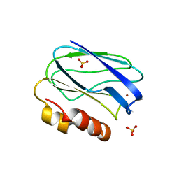 | | Three-dimensional structure of the mutant K109A of Paracoccus pantotrophus pseudoazurin at pH 7.0 | | 分子名称: | COPPER (II) ION, PSEUDOAZURIN, SULFATE ION | | 著者 | Freire, F, Mestre, A, Pinho, J, Najmudin, S, Bonifacio, C, Pauleta, S.R, Romao, M.J. | | 登録日 | 2013-07-15 | | 公開日 | 2014-07-30 | | 最終更新日 | 2023-12-20 | | 実験手法 | X-RAY DIFFRACTION (1.76 Å) | | 主引用文献 | Exploring the Surface Determinants of Paracoccus Pantotrophus Pseudoazurin
To be Published
|
|
4BWU
 | | Three-dimensional structure of the K109A mutant of Paracoccus pantotrophus pseudoazurin at pH 5.5 | | 分子名称: | COPPER (II) ION, PSEUDOAZURIN, SULFATE ION | | 著者 | Freire, F, Mestre, A, Pinho, J, Najmudin, S, Bonifacio, C, Pauleta, S.R, Romao, M.J. | | 登録日 | 2013-07-04 | | 公開日 | 2014-07-16 | | 最終更新日 | 2023-12-20 | | 実験手法 | X-RAY DIFFRACTION (1.76 Å) | | 主引用文献 | Exploring the Surface Determinants of Paracoccus Pantotrophus Pseudoazurin
To be Published
|
|
4BWT
 | | Three-dimensional structure of Paracoccus pantotrophus pseudoazurin at pH 6.5 | | 分子名称: | COPPER (II) ION, PSEUDOAZURIN, SULFATE ION | | 著者 | Freire, F, Mestre, A, Pinho, J, Najmudin, S, Bonifacio, C, Pauleta, S.R, Romao, M.J. | | 登録日 | 2013-07-04 | | 公開日 | 2014-07-16 | | 最終更新日 | 2023-12-20 | | 実験手法 | X-RAY DIFFRACTION (1.76 Å) | | 主引用文献 | Exploring the Surface Determinants of Paracoccus Pantotrophus Pseudoazurin
To be Published
|
|
4AB4
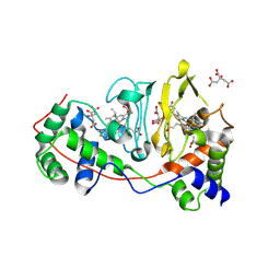
 | | Structure of Xenobiotic Reductase B from Pseudomonas putida in complex with TNT | | 分子名称: | 1,2-ETHANEDIOL, 2,4,6-TRINITROTOLUENE, FLAVIN MONONUCLEOTIDE, ... | | 著者 | Carvalho, A.L, Mukhopadhyay, A, Romao, M.J, Bursakov, S, Kladova, A, Ramos, J.L, van Dillewijn, P. | | 登録日 | 2011-12-06 | | 公開日 | 2012-12-19 | | 最終更新日 | 2023-12-20 | | 実験手法 | X-RAY DIFFRACTION (1.5 Å) | | 主引用文献 | Structural Determinants of Aromatic Ring Reduction in the Xenobiotic Reductase B from Pseudomonas Putida
To be Published
|
|
4AEO
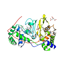
 | | Structure of Xenobiotic Reductase B from Pseudomonas putida in complex with TNT | | 分子名称: | 2,4,6-TRINITROTOLUENE, FLAVIN MONONUCLEOTIDE, GLYCEROL, ... | | 著者 | Carvalho, A.L, Mukhopadhyay, A, Romao, M.J, Bursakov, S, Kladova, A, Ramos, J.L, van Dillewijn, P. | | 登録日 | 2012-01-11 | | 公開日 | 2013-01-30 | | 最終更新日 | 2024-05-08 | | 実験手法 | X-RAY DIFFRACTION (1.7 Å) | | 主引用文献 | Structural Determinants of Aromatic Ring Reduction in the Xenobiotic Reductase B from Pseudomonas Putida
To be Published
|
|
2P1E
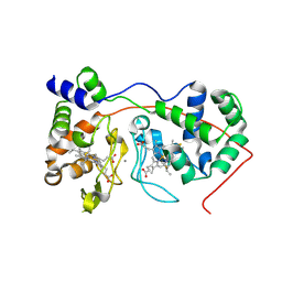
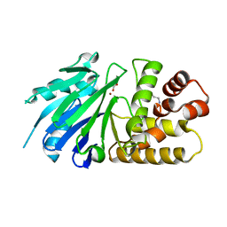 | | Crystal structure of the Leishmania infantum glyoxalase II with D-Lactate at the active site | | 分子名称: | Glyoxalase II, LACTIC ACID, SPERMIDINE, ... | | 著者 | Trincao, J, Barata, L, Najmudin, S, Bonifacio, C, Romao, M.J. | | 登録日 | 2007-03-05 | | 公開日 | 2008-01-15 | | 最終更新日 | 2023-11-15 | | 実験手法 | X-RAY DIFFRACTION (1.9 Å) | | 主引用文献 | Catalysis and Structural Properties of Leishmania infantum Glyoxalase II: Trypanothione Specificity and Phylogeny.
Biochemistry, 47, 2008
|
|
2P18
 | | Crystal structure of the Leishmania infantum glyoxalase II | | 分子名称: | ACETIC ACID, Glyoxalase II, SPERMIDINE, ... | | 著者 | Trincao, J, Barata, L, Najmudin, S, Bonifacio, C, Romao, M.J. | | 登録日 | 2007-03-02 | | 公開日 | 2008-01-15 | | 最終更新日 | 2023-08-30 | | 実験手法 | X-RAY DIFFRACTION (1.8 Å) | | 主引用文献 | Catalysis and Structural Properties of Leishmania infantum Glyoxalase II: Trypanothione Specificity and Phylogeny.
Biochemistry, 47, 2008
|
|


